

360
10 Proteins of Muscle and the Cytoskeleton
case, two different polypeptides are produced by
alternative splicing from one lamin gene. In flies,
a further protein associated with the inner nuclear membrane, otefin (45 kDa) , shows no similarity to lamins or to other IF proteins [126, 210].
10.11 Further Intracellular Structural
Proteins
Various proteins associated with the cell plasma
membrane link the cytoskeleton with integral
membrane proteins and are very important for
attachment of the cell to various surfaces, for cell
movement and for the structural organization of
the cell surface. The most easily approachable
system, and therefore the most investigated, is
that of isolated membranes of vertebrate erythrocytes (erythrocyte ghosts). Erythrocytes show
extraordinary mechanical stability; during their
lifetime of 120 days, human erythrocytes are
forced through the capillary bed 200000 times
and are radically deformed in the process. As
they forfeit most of their cytoskeleton during differentiation, their mechanical strength depends
largely on the structure of the cell membrane
[42]. The central role amongst the structural proteins of the erythrocyte membrane is taken by
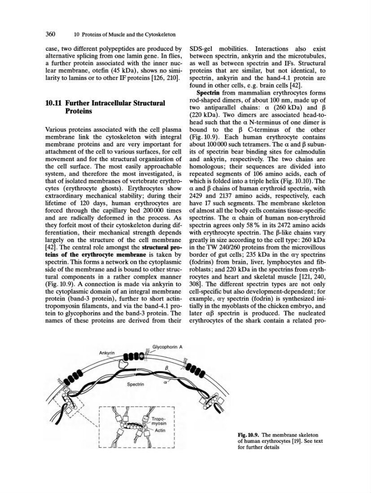
spectrin. This forms a network on the cytoplasmic
side of the membrane and is bound to other structural components in a rather complex manner
(Fig. 10.9). A connection is made via ankyrin to
the cytoplasmic domain of an integral membrane
protein (band-3 protein), further to short actintropomyosin filaments, and via the band-4.1 protein to glycophorins and the band-3 protein. The
names of these proteins are derived from their
SDS-gel mobilities. Interactions also exist
between spectrin, ankyrin and the microtubules,
as well as between spectrin and IFs. Structural
proteins that are similar, but not identical, to
spectrin, ankyrin and the hand-4.1 protein are
found in other cells, e.g. brain cells [42].
Spectrin from mammalian erythrocytes forms
rod-shaped dimers, of about 100 nm, made up of
two antiparallel chains: a (260 kDa) and ~
(220 kDa). Two dimers are associated head-tohead such that the a N-terminus of one dimer is
bound to the ~ C-terminus of the other
(Fig. 10.9). Each human erythrocyte contains
about 100000 such tetramers. The a and ~ subunits of spectrin bear binding sites for calmodulin
and ankyrin, respectively. The two chains are
homologous; their sequences are divided into
repeated segments of 106 amino acids, each of
which is folded into a triple helix (Fig. 10.10). The
a and ~ chains of human erythroid spectrin, with
2429 and 2137 amino acids, respectively, each
have 17 such segments. The membrane skeleton
of almost all the body cells contains tissue-specific
spectrins. The a chain of human non-erythroid
spectrin agrees only 58 % in its 2472 amino acids
with erythrocyte spectrin. The ~-like chains vary
greatly in size according to the cell type: 260 kDa
in the TW 240/260 proteins from the microvillous
border of gut cells; 235 kDa in the ay spectrins
(fodrins) from brain, liver, lymphocytes and fibroblasts; and 220 kDa in the spectrins from erythrocytes and heart and skeletal muscle [121,240,
308]. The different spectrin types are not only
cell-specific but also development-dependent; for
example, ay spectrin (fodrin) is synthesized initially in the myoblasts of the chicken embryo, and
later a~ spectrin is produced. The nucleated
erythrocytes of the shark contain a related proFig.l0.9. The membrane skeleton
of human erythrocytes [19]. See text
for further details
10 Proteins of Muscle and the Cytoskeleton
case, two different polypeptides are produced by
alternative splicing from one lamin gene. In flies,
a further protein associated with the inner nuclear membrane, otefin (45 kDa) , shows no similarity to lamins or to other IF proteins [126, 210].
10.11 Further Intracellular Structural
Proteins
Various proteins associated with the cell plasma
membrane link the cytoskeleton with integral
membrane proteins and are very important for
attachment of the cell to various surfaces, for cell
movement and for the structural organization of
the cell surface. The most easily approachable
system, and therefore the most investigated, is
that of isolated membranes of vertebrate erythrocytes (erythrocyte ghosts). Erythrocytes show
extraordinary mechanical stability; during their
lifetime of 120 days, human erythrocytes are
forced through the capillary bed 200000 times
and are radically deformed in the process. As
they forfeit most of their cytoskeleton during differentiation, their mechanical strength depends
largely on the structure of the cell membrane
[42]. The central role amongst the structural proteins of the erythrocyte membrane is taken by
spectrin. This forms a network on the cytoplasmic
side of the membrane and is bound to other structural components in a rather complex manner
(Fig. 10.9). A connection is made via ankyrin to
the cytoplasmic domain of an integral membrane
protein (band-3 protein), further to short actintropomyosin filaments, and via the band-4.1 protein to glycophorins and the band-3 protein. The
names of these proteins are derived from their
SDS-gel mobilities. Interactions also exist
between spectrin, ankyrin and the microtubules,
as well as between spectrin and IFs. Structural
proteins that are similar, but not identical, to
spectrin, ankyrin and the hand-4.1 protein are
found in other cells, e.g. brain cells [42].
Spectrin from mammalian erythrocytes forms
rod-shaped dimers, of about 100 nm, made up of
two antiparallel chains: a (260 kDa) and ~
(220 kDa). Two dimers are associated head-tohead such that the a N-terminus of one dimer is
bound to the ~ C-terminus of the other
(Fig. 10.9). Each human erythrocyte contains
about 100000 such tetramers. The a and ~ subunits of spectrin bear binding sites for calmodulin
and ankyrin, respectively. The two chains are
homologous; their sequences are divided into
repeated segments of 106 amino acids, each of
which is folded into a triple helix (Fig. 10.10). The
a and ~ chains of human erythroid spectrin, with
2429 and 2137 amino acids, respectively, each
have 17 such segments. The membrane skeleton
of almost all the body cells contains tissue-specific
spectrins. The a chain of human non-erythroid
spectrin agrees only 58 % in its 2472 amino acids
with erythrocyte spectrin. The ~-like chains vary
greatly in size according to the cell type: 260 kDa
in the TW 240/260 proteins from the microvillous
border of gut cells; 235 kDa in the ay spectrins
(fodrins) from brain, liver, lymphocytes and fibroblasts; and 220 kDa in the spectrins from erythrocytes and heart and skeletal muscle [121,240,
308]. The different spectrin types are not only
cell-specific but also development-dependent; for
example, ay spectrin (fodrin) is synthesized initially in the myoblasts of the chicken embryo, and
later a~ spectrin is produced. The nucleated
erythrocytes of the shark contain a related proFig.l0.9. The membrane skeleton
of human erythrocytes [19]. See text
for further details