

line receptor of the skeletal muscles, as in the
case of D-tubocurarine. With dissociation constants of 10- 10 to 10- 11 molll, the neurotoxins bind
more tightly than curare itself and are therefore
useful tools for the isolation of the receptors.
Other neurotoxins act presynaptically, disturbing
the release of acetylcholine; their toxicity is
greater than that of the postsynaptic variety. Poisonous effects on the heart (cardiotoxic effects)
and the lysis of erythrocytes or other cells (cytotoxic effects) are brought about by further toxins
that apparently act primarily on the cell membrane and are, therefore, known as membrane
toxins. The haemolytic and toxic effects of these
venom components are markedly increased in the
presence of phospholipase. The snake venom toxins are referred to either by the initials of the snake's name with additional numbers or letters (e.g.
"Lc-a" from Laticauda colubrina) , or by colourful
names such as "cobrotoxin" (from Naja naja
atra), "erabutoxin" from Laticauda semifasciata)
or "bungarotoxin" (from Bungarus multicinctus).
Approximately 150 snake venom toxins have so
far been sequenced [9, 13] and in several cases
the spatial structure has also been determined
[11]. Two classes are distinguishable on the basis
of structure and activity spectrum, viz. the
postsynaptic-acting neurotoxins and membrane
toxins and the presynaptic neurotoxins.
Numerous postsynaptic-acting neurotoxins are
known from the cobras and the sea snakes, and
the membrane toxins are known mostly from the
cobras. They are all polypeptides of 60-74 amino
acids with four or five disulphide bridges, and
apparently all arose from a common ancestral
form with about 60 amino acids [9, 13, 51]. Several venom components belonging to this protein
family show only very limited toxicity [26]. The
neurotoxins may be divided into "short", with
60-52 amino acids and four disulphide bridges,
and "long", with 71-74 amino acids and five disulphide bridges. However, several neurotoxins are
found in Laticauda species with 66 amino acids
and five disulphide bridges, or with 69 amino
acids and only four disulphide bridges [35, 36,
48]. The membrane toxins all have about 60
amino acids. The sequences of the postsynapticacting neurotoxins and the membrane toxins contain ten invariant positions; considering the neurotoxins alone, there are in fact 22 conserved
positions, of which only four or five are essential
for the toxicity. one characteristic of these
sequences is that two, or even three, identical
amino acids are often found next to each other;
five to ten such homodipeptides are consistently
9.1.1 The Toxins of Snake Venoms
321
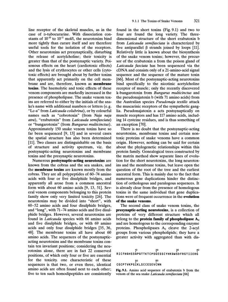
found in the short toxins (Fig.9.1) and two to
four are found the long variety. The threedimensional structure of the short erabutoxin b
from Laticauda semifasciata is characterized by
five antiparallel 13 strands joined by loops [11].
Relatively little is known about the biosynthesis
of the snake venom toxins; however, the precursor of the erabutoxin a from the poison gland of
Laticauda fasciata has been sequenced via the
cDNA and consists only of a 21-amino-acid signal
sequence and the sequence of the mature toxin
[66]. Most of the postsynaptic-acting neurotoxins
bind specifically to the nicotinic acetylcholine
receptor of muscle; only the recently discovered
k-bungarotoxin from Bungarus multicinctus and
the pseudonajatoxin b (with 71 amino acids) from
the Australian species Pseudonaja textilis attack
the muscarinic receptors of the sympathetic ganglia. Pseudonajatoxin a acts postsynaptically on
muscle receptors and has 117 amino acids, including 14 cysteine residues, and is thus something of
an exception [70].
There is no doubt that the postsynaptic-acting
neurotoxins, membrane toxins and certain nontoxic proteins of snake venoms have a common
origin. However, nothing can be said for certain
about the phylogenetic relationships within this
protein family. Genealogical trees constructed by
the matrix method show separate lines of evolution for the short neurotoxins, the long neurotoxins and the membrane toxins, but leave open the
question of the root of the tree and the earliest
ancestral form. This is mainly due to the fact that
numerous gene duplications hinder the distinction of orthologous and paralogous sequences. It
is already clear from the presence of homologous
toxins in the same individual that gene duplications were of frequent occurrence in the evolution
of the snake venoms.
The second class of snake venom toxins, the
presynaptic-acting neurotoxins, is a collection of
proteins of very different structure which all
belong to the protein family of phospholipase A2
and are homologous to the corresponding enzyme
proteins. Phospholipases A2 cleave the 2-acyl
groups from various phospholipids; they have a
greater activity with aggregated than with dis~
~
~
~
RICFNQHSSQPQTTKTCPSGESSCYHKQWSDFRGTIIERG
?o
~o
CGCPTVKPGIKLSCCESEVCNN
Fig.9.1. Amino acid sequence of erabutoxin b from the
venom of the sea snake Laticauda semifasciata [66]
case of D-tubocurarine. With dissociation constants of 10- 10 to 10- 11 molll, the neurotoxins bind
more tightly than curare itself and are therefore
useful tools for the isolation of the receptors.
Other neurotoxins act presynaptically, disturbing
the release of acetylcholine; their toxicity is
greater than that of the postsynaptic variety. Poisonous effects on the heart (cardiotoxic effects)
and the lysis of erythrocytes or other cells (cytotoxic effects) are brought about by further toxins
that apparently act primarily on the cell membrane and are, therefore, known as membrane
toxins. The haemolytic and toxic effects of these
venom components are markedly increased in the
presence of phospholipase. The snake venom toxins are referred to either by the initials of the snake's name with additional numbers or letters (e.g.
"Lc-a" from Laticauda colubrina) , or by colourful
names such as "cobrotoxin" (from Naja naja
atra), "erabutoxin" from Laticauda semifasciata)
or "bungarotoxin" (from Bungarus multicinctus).
Approximately 150 snake venom toxins have so
far been sequenced [9, 13] and in several cases
the spatial structure has also been determined
[11]. Two classes are distinguishable on the basis
of structure and activity spectrum, viz. the
postsynaptic-acting neurotoxins and membrane
toxins and the presynaptic neurotoxins.
Numerous postsynaptic-acting neurotoxins are
known from the cobras and the sea snakes, and
the membrane toxins are known mostly from the
cobras. They are all polypeptides of 60-74 amino
acids with four or five disulphide bridges, and
apparently all arose from a common ancestral
form with about 60 amino acids [9, 13, 51]. Several venom components belonging to this protein
family show only very limited toxicity [26]. The
neurotoxins may be divided into "short", with
60-52 amino acids and four disulphide bridges,
and "long", with 71-74 amino acids and five disulphide bridges. However, several neurotoxins are
found in Laticauda species with 66 amino acids
and five disulphide bridges, or with 69 amino
acids and only four disulphide bridges [35, 36,
48]. The membrane toxins all have about 60
amino acids. The sequences of the postsynapticacting neurotoxins and the membrane toxins contain ten invariant positions; considering the neurotoxins alone, there are in fact 22 conserved
positions, of which only four or five are essential
for the toxicity. one characteristic of these
sequences is that two, or even three, identical
amino acids are often found next to each other;
five to ten such homodipeptides are consistently
9.1.1 The Toxins of Snake Venoms
321
found in the short toxins (Fig.9.1) and two to
four are found the long variety. The threedimensional structure of the short erabutoxin b
from Laticauda semifasciata is characterized by
five antiparallel 13 strands joined by loops [11].
Relatively little is known about the biosynthesis
of the snake venom toxins; however, the precursor of the erabutoxin a from the poison gland of
Laticauda fasciata has been sequenced via the
cDNA and consists only of a 21-amino-acid signal
sequence and the sequence of the mature toxin
[66]. Most of the postsynaptic-acting neurotoxins
bind specifically to the nicotinic acetylcholine
receptor of muscle; only the recently discovered
k-bungarotoxin from Bungarus multicinctus and
the pseudonajatoxin b (with 71 amino acids) from
the Australian species Pseudonaja textilis attack
the muscarinic receptors of the sympathetic ganglia. Pseudonajatoxin a acts postsynaptically on
muscle receptors and has 117 amino acids, including 14 cysteine residues, and is thus something of
an exception [70].
There is no doubt that the postsynaptic-acting
neurotoxins, membrane toxins and certain nontoxic proteins of snake venoms have a common
origin. However, nothing can be said for certain
about the phylogenetic relationships within this
protein family. Genealogical trees constructed by
the matrix method show separate lines of evolution for the short neurotoxins, the long neurotoxins and the membrane toxins, but leave open the
question of the root of the tree and the earliest
ancestral form. This is mainly due to the fact that
numerous gene duplications hinder the distinction of orthologous and paralogous sequences. It
is already clear from the presence of homologous
toxins in the same individual that gene duplications were of frequent occurrence in the evolution
of the snake venoms.
The second class of snake venom toxins, the
presynaptic-acting neurotoxins, is a collection of
proteins of very different structure which all
belong to the protein family of phospholipase A2
and are homologous to the corresponding enzyme
proteins. Phospholipases A2 cleave the 2-acyl
groups from various phospholipids; they have a
greater activity with aggregated than with dis~
~
~
~
RICFNQHSSQPQTTKTCPSGESSCYHKQWSDFRGTIIERG
?o
~o
CGCPTVKPGIKLSCCESEVCNN
Fig.9.1. Amino acid sequence of erabutoxin b from the
venom of the sea snake Laticauda semifasciata [66]