

The majority of mollusc haemocyanins consist
of only one type of subunit, but electrophoretically or immunologically distinct types are found
in some species. The 100S haemocyanin of the
snail Murex fulvescens is made up of ten each of
two subunits, A and B, which differ especially in
their respiratory properties. Reassociation products with the characteristics of the native molecule result only from mixtures of the two subunit
types; A or B alone produces only dimers. Heterogeneons subunits are also found in Nautilus
pompilius, the snail Megathura crenulata, and the
Polyplacophora [63, 108]. Single subunits, and
even isolated domains, can bind O2 but generally
exhibit no cooperativity and a reduced Bohr
effect. Reassociation of the subunits usually leads
to molecules with the structural and functional
properties of the native haemocyanin [108].
7.4 Haemerythrins
The haemerythrins have a curious, sporadic distribution (Table 7.2). Haemerythrin-containing
cells (erythrocytes) are found in the coelum fluid
of all sipunculids and priapulids as well as several
species of the ecardinate brachiopods (e.g. Lingula reevei) , and are present in the enucleate
blood cells and muscles of the polychaete MageZona papillicornis. Blood cells and muscles of the
sipunculids also contain haemerythrins with
somewhat different properties (myohaemerythrins). The myohaemerythrins are monomers of
about 13.S kDa, whereas the pigments in the
blood and coelom cells of the sipunculids, priapulids and brachiopods are trimers, tetramers or
octomers of subunits with the same size [83, 108,
174]. The major vascular haemerythrin of MageZona has the unusually large size for an intracellular respiratory pigment of 693 ± 32 kDa [113].
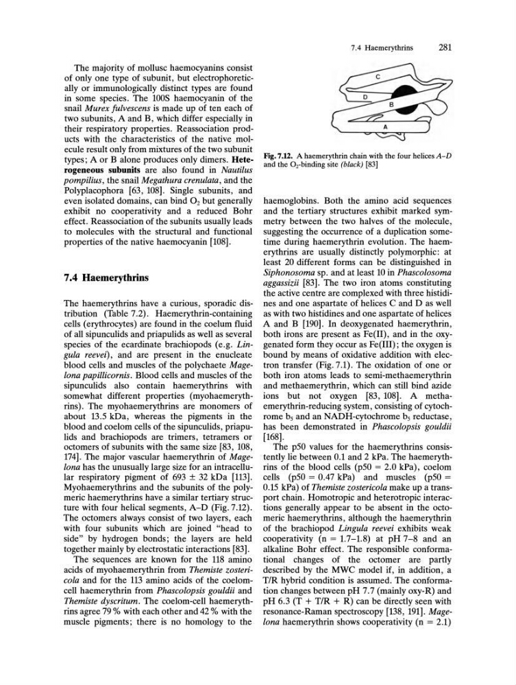
Myohaemerythrins and the subunits of the polymeric haemerythrins have a similar tertiary structure with four helical segments, A-D (Pig. 7.12).
The octomers always consist of two layers, each
with four subunits which are joined "head to
side" by hydrogen bonds; the layers are held
together mainly by electrostatic interactions [83].
The sequences are known for the 118 amino
acids of myohaemerythrin from Themiste zostericola and for the 113 amino acids of the coelomcell haemerythrin from Phascolopsis gouldii and
Themiste dyscritum. The coelom-cell haemerythrins agree 79 % with each other and 42 % with the
muscle pigments; there is no homology to the
7.4 Haemerythrins
281
Fig.7.12. A haemerythrin chain with the four helices A-D
and the Oz-binding site (black) [83]
haemoglobins. Both the amino acid sequences
and the tertiary structures exhibit marked symmetry between the two halves of the molecule,
suggesting the occurrence of a duplication sometime during haemerythrin evolution. The haemerythrins are usually distinctly polymorphic: at
least 20 different forms can be distinguished in
Siphonosoma sp. and at least 10 in PhascoZosoma
aggassizii [83]. The two iron atoms constituting
the active centre are complexed with three histidines and one aspartate of helices C and D as well
as with two histidines and one aspartate of helices
A and B [190]. In deoxygenated haemerythrin,
both irons are present as Pe(II), and in the oxygenated form they occur as Pe(III); the oxygen is
bound by means of oxidative addition with electron transfer (Pig. 7.1). The oxidation of one or
both iron atoms leads to semi-methaemerythrin
and methaemerythrin, which can still bind azide
ions but not oxygen [83, 108]. A methaemerythrin-reducing system, consisting of cytochrome bs and an NADH-cytochrome bs reductase,
has been demonstrated in PhascoZopsis gouldii
[168].
The pSO values for the haemerythrins consistently lie between 0.1 and 2 kPa. The haemerythrins of the blood cells (pSO = 2.0 kPa), coelom
cells (pSO = 0.47 kPa) and muscles (pSO =
O.lS kPa) of Themiste zostericoZa make up a transport chain. Homotropic and heterotropic interactions generally appear to be absent in the octomeric haemerythrins, although the haemerythrin
of the brachiopod Lingula reevei exhibits weak
cooperativity (n = 1.7-1.8) at pH 7-8 and an
alkaline Bohr effect. The responsible conformational changes of the octomer are partly
described by the MWC model if, in addition, a
TIR hybrid condition is assumed. The conformation changes between pH 7.7 (mainly oxy-R) and
pH 6.3 (T + T/R + R) can be directly seen with
resonance-Raman spectroscopy [138, 191]. Magelona haemerythrin shows cooperativity (n = 2.1)
of only one type of subunit, but electrophoretically or immunologically distinct types are found
in some species. The 100S haemocyanin of the
snail Murex fulvescens is made up of ten each of
two subunits, A and B, which differ especially in
their respiratory properties. Reassociation products with the characteristics of the native molecule result only from mixtures of the two subunit
types; A or B alone produces only dimers. Heterogeneons subunits are also found in Nautilus
pompilius, the snail Megathura crenulata, and the
Polyplacophora [63, 108]. Single subunits, and
even isolated domains, can bind O2 but generally
exhibit no cooperativity and a reduced Bohr
effect. Reassociation of the subunits usually leads
to molecules with the structural and functional
properties of the native haemocyanin [108].
7.4 Haemerythrins
The haemerythrins have a curious, sporadic distribution (Table 7.2). Haemerythrin-containing
cells (erythrocytes) are found in the coelum fluid
of all sipunculids and priapulids as well as several
species of the ecardinate brachiopods (e.g. Lingula reevei) , and are present in the enucleate
blood cells and muscles of the polychaete MageZona papillicornis. Blood cells and muscles of the
sipunculids also contain haemerythrins with
somewhat different properties (myohaemerythrins). The myohaemerythrins are monomers of
about 13.S kDa, whereas the pigments in the
blood and coelom cells of the sipunculids, priapulids and brachiopods are trimers, tetramers or
octomers of subunits with the same size [83, 108,
174]. The major vascular haemerythrin of MageZona has the unusually large size for an intracellular respiratory pigment of 693 ± 32 kDa [113].
Myohaemerythrins and the subunits of the polymeric haemerythrins have a similar tertiary structure with four helical segments, A-D (Pig. 7.12).
The octomers always consist of two layers, each
with four subunits which are joined "head to
side" by hydrogen bonds; the layers are held
together mainly by electrostatic interactions [83].
The sequences are known for the 118 amino
acids of myohaemerythrin from Themiste zostericola and for the 113 amino acids of the coelomcell haemerythrin from Phascolopsis gouldii and
Themiste dyscritum. The coelom-cell haemerythrins agree 79 % with each other and 42 % with the
muscle pigments; there is no homology to the
7.4 Haemerythrins
281
Fig.7.12. A haemerythrin chain with the four helices A-D
and the Oz-binding site (black) [83]
haemoglobins. Both the amino acid sequences
and the tertiary structures exhibit marked symmetry between the two halves of the molecule,
suggesting the occurrence of a duplication sometime during haemerythrin evolution. The haemerythrins are usually distinctly polymorphic: at
least 20 different forms can be distinguished in
Siphonosoma sp. and at least 10 in PhascoZosoma
aggassizii [83]. The two iron atoms constituting
the active centre are complexed with three histidines and one aspartate of helices C and D as well
as with two histidines and one aspartate of helices
A and B [190]. In deoxygenated haemerythrin,
both irons are present as Pe(II), and in the oxygenated form they occur as Pe(III); the oxygen is
bound by means of oxidative addition with electron transfer (Pig. 7.1). The oxidation of one or
both iron atoms leads to semi-methaemerythrin
and methaemerythrin, which can still bind azide
ions but not oxygen [83, 108]. A methaemerythrin-reducing system, consisting of cytochrome bs and an NADH-cytochrome bs reductase,
has been demonstrated in PhascoZopsis gouldii
[168].
The pSO values for the haemerythrins consistently lie between 0.1 and 2 kPa. The haemerythrins of the blood cells (pSO = 2.0 kPa), coelom
cells (pSO = 0.47 kPa) and muscles (pSO =
O.lS kPa) of Themiste zostericoZa make up a transport chain. Homotropic and heterotropic interactions generally appear to be absent in the octomeric haemerythrins, although the haemerythrin
of the brachiopod Lingula reevei exhibits weak
cooperativity (n = 1.7-1.8) at pH 7-8 and an
alkaline Bohr effect. The responsible conformational changes of the octomer are partly
described by the MWC model if, in addition, a
TIR hybrid condition is assumed. The conformation changes between pH 7.7 (mainly oxy-R) and
pH 6.3 (T + T/R + R) can be directly seen with
resonance-Raman spectroscopy [138, 191]. Magelona haemerythrin shows cooperativity (n = 2.1)