

addition to 6-mers and 12-mers are reported to
occur in the crayfish Cherax destructor. The 6mers are the rule in the Palinura (Panulirus, Scyllarus) , Natantia (Palaemon) and Euphausiacea
[108, 115, 178]. The haemocyanins of most Chelicerata are larger than those of the Crustacea, with
only the Xiphosura (Limulus, Tachypleus) having
48-mers, the scorpions, uropygi, amblipygi and
most spiders having 24-mers, and the opiliones
having 12-mers [16, 108, 115]. Some spider species have haemocyanin molecules smaller than
24-mer: Cupiennius salei has 12-mers, and 6-mers
occur in Filistata insidiatrix and Dysdera crocata
[108, 115]. The haemocyanin of the chilopod Scutigera coleoptrata is apparently made up of six
hexamers [108].
The dissociation of native haemocyanins into
subunits is a slow process which is promoted at
alkaline pH, and strongly promoted by the
removal of divalent cations and the addition of
denaturing reagents. Thus, dissociation of haemocyanin in Limulus and other chelicerates is
initiated only at pH 9.6 in the presence of Ca z +,
but at pH 8.8 upon addition of 10 mM EDTA or
2 % SDS + 1 % mercaptoethanol. Some crustacean haemocyanins can resist dissociation at
pH 10 in the presence of Ca z +. Reaggregation at
neutral pH in the presence of a sufficiently high
Ca z + concentration is often incomplete or results
in products which are not identical to the native
haemocyanin [77, 108]. The subunits of the arthropod haemocyanins are polypeptides with about
630-670 amino acids, 20 a-helices and a sevenstranded ~ structure; they are subdivided into
three domains, the middle domain bearing the
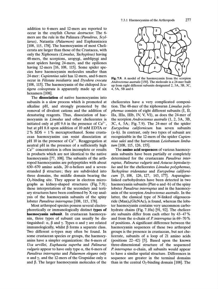
Oz-binding site. They appear in electron micrographs as kidney-shaped structures (Fig. 7.9);
these interpretations of the secondary and tertiary structures have been confirmed by X-ray analysis of the haemocyanin subunits of the spiny
lobster Panulirus interruptus [108, 115, 178].
Most arthropod species possess several electrophoretically or immunologically distinct types of
baemocyanin subunit. In crustacean haemocyanin, three types of subunit can usually be distinguished: a, ~ and y. Types a and yare related
immunologically, whilst ~ forms a separate class.
Two different a-types may often be found. In
some crustacean species or groups, the haemocyanins have a simpler organization: the 6-mers of
Uca urvillei, Euphausia superba and Palinurus
vulgaris appear to have only type a, the 6-mers of
Panulirus interruptus and Palaemon elegans only
a and y, and the 12-mers of the Grapsidae only a
and ~. The larger haemocyanin molecules of the
7.3.1 Haemocyanins of the Arthropods
277
Fig.7.9. A model of the haemocyanin from the scorpion
Androctonus australis [150]. The molecule is a 24-mer built
up from eight different subunits designated 2, 3A, 3B, 3C,
4, SA, 5B and 6
chelicerates have a very complicated composition. The 48-mer of the xiphosuran Limulus polyphemus consists of eight different subunits (I, II,
IIa, IlIa, IIIb, IV, V, VI), as does the 24-mer of
the scorpion Androctonus australis (1,2, 3A, 3B,
3C,4, 5A; Fig. 7.9). The 24-mer of the spider
Eurypelma californicum has seven subunits
(a-h). In contrast, only two types of subunit are
recognizable in the 12-mers of the spider Cupiennius salei and the harvestman Leiobunum limbatum [108, 115, 126, 155].
The amino acid sequences of various haemocyanin subunits have been partially or completely
determined for the crustaceans Panulirus interruptus, Palinurus vulgaris and Astacus leptodactyIus and for the chelicerates Limulus polyphemus,
Tachypleus tridentatus and Eurypelma californicum [5, 108, 126, 127, 143, 177]. Asparaginelinked carbohydrates have been detected in two
haemocyanin subunits (Pint-a and -b) of the spiny
lobster Panulirus interruptus and in the haemocyanin of the scorpion Androctonus australis. In the
latter, the classical type of N-linked oligo saccaride (ManMGlcNAc)z is found, whereas the lobster haemocyanin contains very uncommon carbohydrate chains (Fig.7.lOa) [91, 92]. The chelicerate subunits differ from each other by 43-47 %
and from the a-chain of P. interruptus in 69-70 %
of positions. A significant difference between the
haemocyanin sequences of these two arthropod
groups is the presence in crustacean, but not chelicerate, subunits of a loop of 21 amino acids
(positions 22-42) [5]. Based upon the known
three-dimensional structure of the sequenced
P. interruptus a-chain, all subunits would appear
to have a similar spatial structure. Differences in
sequence are greater in the terminal domains
than in the central Oz-binding domain [108]. The
occur in the crayfish Cherax destructor. The 6mers are the rule in the Palinura (Panulirus, Scyllarus) , Natantia (Palaemon) and Euphausiacea
[108, 115, 178]. The haemocyanins of most Chelicerata are larger than those of the Crustacea, with
only the Xiphosura (Limulus, Tachypleus) having
48-mers, the scorpions, uropygi, amblipygi and
most spiders having 24-mers, and the opiliones
having 12-mers [16, 108, 115]. Some spider species have haemocyanin molecules smaller than
24-mer: Cupiennius salei has 12-mers, and 6-mers
occur in Filistata insidiatrix and Dysdera crocata
[108, 115]. The haemocyanin of the chilopod Scutigera coleoptrata is apparently made up of six
hexamers [108].
The dissociation of native haemocyanins into
subunits is a slow process which is promoted at
alkaline pH, and strongly promoted by the
removal of divalent cations and the addition of
denaturing reagents. Thus, dissociation of haemocyanin in Limulus and other chelicerates is
initiated only at pH 9.6 in the presence of Ca z +,
but at pH 8.8 upon addition of 10 mM EDTA or
2 % SDS + 1 % mercaptoethanol. Some crustacean haemocyanins can resist dissociation at
pH 10 in the presence of Ca z +. Reaggregation at
neutral pH in the presence of a sufficiently high
Ca z + concentration is often incomplete or results
in products which are not identical to the native
haemocyanin [77, 108]. The subunits of the arthropod haemocyanins are polypeptides with about
630-670 amino acids, 20 a-helices and a sevenstranded ~ structure; they are subdivided into
three domains, the middle domain bearing the
Oz-binding site. They appear in electron micrographs as kidney-shaped structures (Fig. 7.9);
these interpretations of the secondary and tertiary structures have been confirmed by X-ray analysis of the haemocyanin subunits of the spiny
lobster Panulirus interruptus [108, 115, 178].
Most arthropod species possess several electrophoretically or immunologically distinct types of
baemocyanin subunit. In crustacean haemocyanin, three types of subunit can usually be distinguished: a, ~ and y. Types a and yare related
immunologically, whilst ~ forms a separate class.
Two different a-types may often be found. In
some crustacean species or groups, the haemocyanins have a simpler organization: the 6-mers of
Uca urvillei, Euphausia superba and Palinurus
vulgaris appear to have only type a, the 6-mers of
Panulirus interruptus and Palaemon elegans only
a and y, and the 12-mers of the Grapsidae only a
and ~. The larger haemocyanin molecules of the
7.3.1 Haemocyanins of the Arthropods
277
Fig.7.9. A model of the haemocyanin from the scorpion
Androctonus australis [150]. The molecule is a 24-mer built
up from eight different subunits designated 2, 3A, 3B, 3C,
4, SA, 5B and 6
chelicerates have a very complicated composition. The 48-mer of the xiphosuran Limulus polyphemus consists of eight different subunits (I, II,
IIa, IlIa, IIIb, IV, V, VI), as does the 24-mer of
the scorpion Androctonus australis (1,2, 3A, 3B,
3C,4, 5A; Fig. 7.9). The 24-mer of the spider
Eurypelma californicum has seven subunits
(a-h). In contrast, only two types of subunit are
recognizable in the 12-mers of the spider Cupiennius salei and the harvestman Leiobunum limbatum [108, 115, 126, 155].
The amino acid sequences of various haemocyanin subunits have been partially or completely
determined for the crustaceans Panulirus interruptus, Palinurus vulgaris and Astacus leptodactyIus and for the chelicerates Limulus polyphemus,
Tachypleus tridentatus and Eurypelma californicum [5, 108, 126, 127, 143, 177]. Asparaginelinked carbohydrates have been detected in two
haemocyanin subunits (Pint-a and -b) of the spiny
lobster Panulirus interruptus and in the haemocyanin of the scorpion Androctonus australis. In the
latter, the classical type of N-linked oligo saccaride (ManMGlcNAc)z is found, whereas the lobster haemocyanin contains very uncommon carbohydrate chains (Fig.7.lOa) [91, 92]. The chelicerate subunits differ from each other by 43-47 %
and from the a-chain of P. interruptus in 69-70 %
of positions. A significant difference between the
haemocyanin sequences of these two arthropod
groups is the presence in crustacean, but not chelicerate, subunits of a loop of 21 amino acids
(positions 22-42) [5]. Based upon the known
three-dimensional structure of the sequenced
P. interruptus a-chain, all subunits would appear
to have a similar spatial structure. Differences in
sequence are greater in the terminal domains
than in the central Oz-binding domain [108]. The