

7.1.2 Ligand Binding and Cooperativity
253
A
B
C
o
a V-LSPADKTNVKAAWGKVGAHAGEYGAEALERMFLSFPTTKTYFPHF-DLS-----HG
~
VHLTPEEKSAVTALWGKV--NVDEVGGEALGRLLVVYPWTQRFFESFGDLSTPDAVMG
E
F
G
a SAQVKGHGKKVADALTNAVAHVDDMPNALSALSDLHAHKLRVDPVNFKLLSHCLLVTLAAH
~
NPKVKAHGKKVLGAFSDGLAHLDNLKGT-FATLSELHCDKLHVDPENFRLLGNVLVCVLAHH
H
a LPAEFTPAVHASLDKFLASVSTVLTSKYR
~
FGKEFTPPVQAAYQKVVAGVANALAHKYH
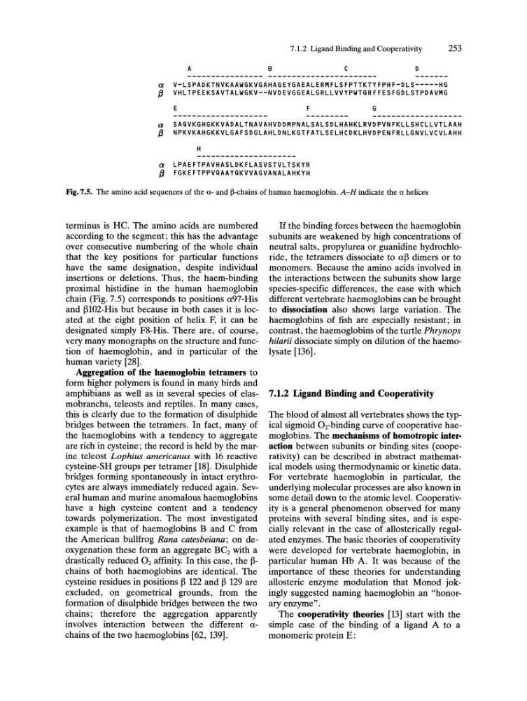
Fig.7.S. The amino acid sequences of the (l- and ~-chains of human haemoglobin. A-H indicate the (l helices
terminus is HC. The amino acids are numbered
according to the segment; this has the advantage
over consecutive numbering of the whole chain
that the key positions for particular functions
have the same designation, despite individual
insertions or deletions. Thus, the haem-binding
proximal histidine in the human haemoglobin
chain (Fig. 7.5) corresponds to positions a97-His
and ~102-His but because in both cases it is located at the eight position of helix F, it can be
designated simply F8-His. There are, of course,
very many monographs on the structure and function of haemoglobin, and in particular of the
human variety [28].
Aggregation of the haemoglobin tetramers to
form higher polymers is found in many birds and
amphibians as well as in several species of elasmobranchs, teleosts and reptiles. In many cases,
this is clearly due to the formation of disulphide
bridges between the tetramers. In fact, many of
the haemoglobins with a tendency to aggregate
are rich in cysteine; the record is held by the marine teleost Lophius americanus with 16 reactive
cysteine-SH groups per tetramer [18]. Disulphide
bridges forming spontaneously in intact erythrocytes are always immediately reduced again. Several human and murine anomalous haemoglobins
have a high cysteine content and a tendency
towards polymerization. The most investigated
example is that of haemoglobins Band C from
the American bullfrog Rana catesbeiana; on deoxygenation these form an aggregate BCz with a
drastically reduced O2 affinity. In this case, the ~chains of both haemoglobins are identical. The
cysteine residues in positions ~ 122 and ~ 129 are
excluded, on geometrical grounds, from the
formation of disulphide bridges between the two
chains; therefore the aggregation apparently
involves interaction between the different achains of the two haemoglobins [62, 139].
If the binding forces between the haemoglobin
subunits are weakened by high concentrations of
neutral salts, propylurea or guanidine hydrochloride, the tetramers dissociate to a~ dimers or to
monomers. Because the amino acids involved in
the interactions between the subunits show large
species-specific differences, the ease with which
different vertebrate haemoglobins can be brought
to dissociation also shows large variation. The
haemoglobins of fish are especially resistant; in
contrast, the haemoglobins of the turtle Phrynops
hilarii dissociate simply on dilution of the haemolysate [136].
7.1.2 Ligand Binding and Cooperativity
The blood of almost all vertebrates shows the typical sigmoid 02-binding curve of cooperative haemoglobins. The mechanisms of homotropic interaction between subunits or binding sites (cooperativity) can be described in abstract mathematical models using thermodynamic or kinetic data.
For vertebrate haemoglobin in particular, the
underlying molecular processes are also known in
some detail down to the atomic level. Cooperativity is a general phenomenon observed for many
proteins with several binding sites, and is especially relevant in the case of allosterically regulated enzymes. The basic theories of cooperativity
were developed for vertebrate haemoglobin, in
particular human Hb A. It was because of the
importance of these theories for understanding
allosteric enzyme modulation that Monod jokingly suggested naming haemoglobin an "honorary enzyme".
The cooperativity theories [13] start with the
simple case of the binding of a ligand A to a
monomeric protein E:
253
A
B
C
o
a V-LSPADKTNVKAAWGKVGAHAGEYGAEALERMFLSFPTTKTYFPHF-DLS-----HG
~
VHLTPEEKSAVTALWGKV--NVDEVGGEALGRLLVVYPWTQRFFESFGDLSTPDAVMG
E
F
G
a SAQVKGHGKKVADALTNAVAHVDDMPNALSALSDLHAHKLRVDPVNFKLLSHCLLVTLAAH
~
NPKVKAHGKKVLGAFSDGLAHLDNLKGT-FATLSELHCDKLHVDPENFRLLGNVLVCVLAHH
H
a LPAEFTPAVHASLDKFLASVSTVLTSKYR
~
FGKEFTPPVQAAYQKVVAGVANALAHKYH
Fig.7.S. The amino acid sequences of the (l- and ~-chains of human haemoglobin. A-H indicate the (l helices
terminus is HC. The amino acids are numbered
according to the segment; this has the advantage
over consecutive numbering of the whole chain
that the key positions for particular functions
have the same designation, despite individual
insertions or deletions. Thus, the haem-binding
proximal histidine in the human haemoglobin
chain (Fig. 7.5) corresponds to positions a97-His
and ~102-His but because in both cases it is located at the eight position of helix F, it can be
designated simply F8-His. There are, of course,
very many monographs on the structure and function of haemoglobin, and in particular of the
human variety [28].
Aggregation of the haemoglobin tetramers to
form higher polymers is found in many birds and
amphibians as well as in several species of elasmobranchs, teleosts and reptiles. In many cases,
this is clearly due to the formation of disulphide
bridges between the tetramers. In fact, many of
the haemoglobins with a tendency to aggregate
are rich in cysteine; the record is held by the marine teleost Lophius americanus with 16 reactive
cysteine-SH groups per tetramer [18]. Disulphide
bridges forming spontaneously in intact erythrocytes are always immediately reduced again. Several human and murine anomalous haemoglobins
have a high cysteine content and a tendency
towards polymerization. The most investigated
example is that of haemoglobins Band C from
the American bullfrog Rana catesbeiana; on deoxygenation these form an aggregate BCz with a
drastically reduced O2 affinity. In this case, the ~chains of both haemoglobins are identical. The
cysteine residues in positions ~ 122 and ~ 129 are
excluded, on geometrical grounds, from the
formation of disulphide bridges between the two
chains; therefore the aggregation apparently
involves interaction between the different achains of the two haemoglobins [62, 139].
If the binding forces between the haemoglobin
subunits are weakened by high concentrations of
neutral salts, propylurea or guanidine hydrochloride, the tetramers dissociate to a~ dimers or to
monomers. Because the amino acids involved in
the interactions between the subunits show large
species-specific differences, the ease with which
different vertebrate haemoglobins can be brought
to dissociation also shows large variation. The
haemoglobins of fish are especially resistant; in
contrast, the haemoglobins of the turtle Phrynops
hilarii dissociate simply on dilution of the haemolysate [136].
7.1.2 Ligand Binding and Cooperativity
The blood of almost all vertebrates shows the typical sigmoid 02-binding curve of cooperative haemoglobins. The mechanisms of homotropic interaction between subunits or binding sites (cooperativity) can be described in abstract mathematical models using thermodynamic or kinetic data.
For vertebrate haemoglobin in particular, the
underlying molecular processes are also known in
some detail down to the atomic level. Cooperativity is a general phenomenon observed for many
proteins with several binding sites, and is especially relevant in the case of allosterically regulated enzymes. The basic theories of cooperativity
were developed for vertebrate haemoglobin, in
particular human Hb A. It was because of the
importance of these theories for understanding
allosteric enzyme modulation that Monod jokingly suggested naming haemoglobin an "honorary enzyme".
The cooperativity theories [13] start with the
simple case of the binding of a ligand A to a
monomeric protein E: