

7 Respiratory Pigments
7.1
7.1.1
7.1.2
7.1.3
7.1.4
7.1.5
7.1.6
7.1.7
7.2
7.2.1
The Haemoglobins and Myoglobins of Vertebrates
The Structure of Haemoglobin
Ligand Binding and Cooperativity
Heterotropic Interactions
Temperature Effects and Adaptations
The Gene Families of Vertebrate Haemoglobins
Myoglobins
Methaemoglobin Formation and Reduction
Invertebrate Haemoglobins and Chlorocruorins
Annelids, Pogonophora and Echiurids
In the animal kingdom there are four types of O2binding (respiratory) pigment with different
structures but very similar functional properties.
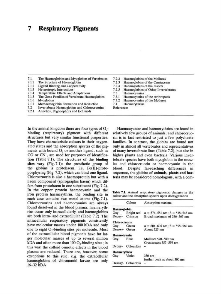
They have characteristic colours in their oxygenated states and the absorption spectra of the pigments with bound O2 or another ligand, such as
CO or CN-, are used for purposes of identification (Table 7.1). The structures of the binding
sites vary (Fig. 7.1): the prosthetic group of
the globins is protohaem, i.e. Fe(II)-protoporphyring (Fig. 7.2), which can bind one ligand.
Chlorocruorin is also a haemoprotein but with a
haem component (spirographis haem) which differs from protohaem in one substituent (Fig. 7.2).
In the copper protein haemocyanin and the
iron protein haemerythrin, the binding site in
each case contains two metal atoms (Fig. 7.1).
Chlorocruorins and haemocyanins are always
found dissolved in the blood plasma; haemerythrins occur only intracellularly, and haemoglobins
are both intra- and extracellular (Table 7.2). The
intracellular respiratory pigments consistently
have molecular masses under 100 kDA and only
one to eight OTbinding sites per molecule. Most
of the extracellular blood pigments have far larger molecular masses of up to several million
kDA and often more than 100 OTbinding sites; in
this way, the colloid osmotic effects in the blood
plasma are reduced. There are, however, some
exceptions to this rule, e.g. the extracellular
haemoglobins of chironomid larvae are only
16-32 kDA.
7.2.2 Haemoglobins of the Molluscs
7.2.3 Haemoglobins of the Crustaceans
7.2.4 Haemoglobins of the Insects
7.2.5 Haemoglobins of Other Invertebrates
7.3
Haemocyanins
7.3.1 Haemocyanins of the Arthropods
7.3.2 Haemocyanins of the Molluscs
7.4
Haemerythrins
References
Haemocyanins and haemerythrins are found in
relatively few groups of animals, and chlorocruorin is in fact restricted to just a few polychaete
families. In contrast, the globins are found not
only in almost all vertebrates and representatives
of many invertebrate lines (Table 7.2), but also in
higher plants and even bacteria. Various invertebrate species have both myoglobin in the muscles and chlorocruorin or haemocyanin in the
blood. Despite far-reaching differences in
sequence, the globins of animals, plants and bacteria may be considered homologous, with a comTable 7.1. Animal respiratory pigments: changes in the
colour and the absorption spectra upon deoxygenation
Colour
Absorption maxima
Haemoglobin
OxyBright red a = 574-581 nm; ~ = 538-545 nm
Deoxy- Crimson
Broad maximum of 556-565 nm
Chlorocmorin
OxyGreen
Desoxy- Green
Haemocyanin
OxyBlue
a = 604-605 nm; ~ = 558-560 nm
About 525 nm
Molluscs 570-580 nm
Crustaceans 557-559 nm
Deoxy- Colourless -
Haemerythrin
OxyViolet
330 nm;
further peak at about 500 nm
Desoxy- Colourless -
7.1
7.1.1
7.1.2
7.1.3
7.1.4
7.1.5
7.1.6
7.1.7
7.2
7.2.1
The Haemoglobins and Myoglobins of Vertebrates
The Structure of Haemoglobin
Ligand Binding and Cooperativity
Heterotropic Interactions
Temperature Effects and Adaptations
The Gene Families of Vertebrate Haemoglobins
Myoglobins
Methaemoglobin Formation and Reduction
Invertebrate Haemoglobins and Chlorocruorins
Annelids, Pogonophora and Echiurids
In the animal kingdom there are four types of O2binding (respiratory) pigment with different
structures but very similar functional properties.
They have characteristic colours in their oxygenated states and the absorption spectra of the pigments with bound O2 or another ligand, such as
CO or CN-, are used for purposes of identification (Table 7.1). The structures of the binding
sites vary (Fig. 7.1): the prosthetic group of
the globins is protohaem, i.e. Fe(II)-protoporphyring (Fig. 7.2), which can bind one ligand.
Chlorocruorin is also a haemoprotein but with a
haem component (spirographis haem) which differs from protohaem in one substituent (Fig. 7.2).
In the copper protein haemocyanin and the
iron protein haemerythrin, the binding site in
each case contains two metal atoms (Fig. 7.1).
Chlorocruorins and haemocyanins are always
found dissolved in the blood plasma; haemerythrins occur only intracellularly, and haemoglobins
are both intra- and extracellular (Table 7.2). The
intracellular respiratory pigments consistently
have molecular masses under 100 kDA and only
one to eight OTbinding sites per molecule. Most
of the extracellular blood pigments have far larger molecular masses of up to several million
kDA and often more than 100 OTbinding sites; in
this way, the colloid osmotic effects in the blood
plasma are reduced. There are, however, some
exceptions to this rule, e.g. the extracellular
haemoglobins of chironomid larvae are only
16-32 kDA.
7.2.2 Haemoglobins of the Molluscs
7.2.3 Haemoglobins of the Crustaceans
7.2.4 Haemoglobins of the Insects
7.2.5 Haemoglobins of Other Invertebrates
7.3
Haemocyanins
7.3.1 Haemocyanins of the Arthropods
7.3.2 Haemocyanins of the Molluscs
7.4
Haemerythrins
References
Haemocyanins and haemerythrins are found in
relatively few groups of animals, and chlorocruorin is in fact restricted to just a few polychaete
families. In contrast, the globins are found not
only in almost all vertebrates and representatives
of many invertebrate lines (Table 7.2), but also in
higher plants and even bacteria. Various invertebrate species have both myoglobin in the muscles and chlorocruorin or haemocyanin in the
blood. Despite far-reaching differences in
sequence, the globins of animals, plants and bacteria may be considered homologous, with a comTable 7.1. Animal respiratory pigments: changes in the
colour and the absorption spectra upon deoxygenation
Colour
Absorption maxima
Haemoglobin
OxyBright red a = 574-581 nm; ~ = 538-545 nm
Deoxy- Crimson
Broad maximum of 556-565 nm
Chlorocmorin
OxyGreen
Desoxy- Green
Haemocyanin
OxyBlue
a = 604-605 nm; ~ = 558-560 nm
About 525 nm
Molluscs 570-580 nm
Crustaceans 557-559 nm
Deoxy- Colourless -
Haemerythrin
OxyViolet
330 nm;
further peak at about 500 nm
Desoxy- Colourless -