

Comparisons of the repeat sequences within and
between species allow conclusions to be drawn
about the frequency with which sequence equalization through horizontal evolution has
occurred; in the case of ubiquitin, it is on average
38 million years since the last such event in mammals, and only 11 million years in Drosophila
[248].
1\vo large, proteolytic complexes are responsible for the degradation of ubiquitin-marked proteins. The 26S complex is about 1500 kDa and in
vitro originates from three smaller subunits
CI-C3. The well-known 650-kDa complex, termed "multicatalytic protease" because of its wide
specificity, or "macropain" or "proteasome", is
identical to the C3 component. This consists of
subunits of 20-35 kDa, organized in a fourringed cylinder, with each ring having up to eight
subunits. The human proteasome in erythrocytes
contains 13 different types of subunit, varying in
both structure and specificity, although these
apparently belong to the same multi-gene family
[155, 202, 255]. The freshly isolated complex is
stimulated by ATP, but this effect diminishes with
storage. Multi-catalytic proteases are apparently
ubiquitous; particles similar to mammalian proteasomes are found in archaebacteria, and they
have been closely studied in lower vertebrates,
insects and crustaceans [44, 59,67, 73, 93, 185].
In the classification of the proteases, one can
distinguish between exopeptidases, which cleave
N- or C-terminal bonds, and endopeptidases,
which attack internal bonds; finer characterization is based upon the structure of the active
centre (Table 3.3). In the absence of information
on amino acid sequences, several characteristic
properties can be used to study the relationship
between the proteases of the lower vertebrates
and invertebrates and those of the mammals;
these include, in particular, inhibition by synthetic and natural inhibitors, the specificity for certain low molecular weight, synthetic substrates,
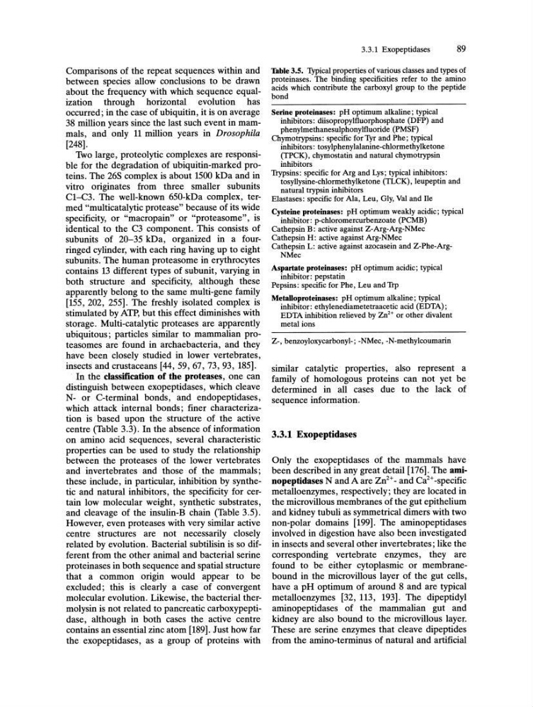
and cleavage of the insulin-B chain (Table 3.5).
However, even proteases with very similar active
centre structures are not necessarily closely
related by evolution. Bacterial subtilisin is so different from the other animal and bacterial serine
proteinases in both sequence and spatial structure
that a common origin would appear to be
excluded; this is clearly a case of convergent
molecular evolution. Likewise, the bacterial thermolysin is not related to pancreatic carboxypeptidase, although in both cases the active centre
contains an essential zinc atom [189]. Just how far
the exopeptidases, as a group of proteins with
3.3.1 Exopeptidases
89
Table 3.5. Typical properties of various classes and types of
proteinases. The binding specificities refer to the amino
acids which contribute the carboxyl group to the peptide
bond
Serine proteinases: pH optimum alkaline; typical
inhibitors: diisopropylfluorphosphate (D FP) and
phenylmethanesulphonylfluoride (PMSF)
Chymotrypsins: specific for Tyr and Phe; typical
inhibitors: tosylphenylalanine-chlormethylketone
(TPCK), chymostatin and natural chymotrypsin
inhibitors
Trypsins: specific for Arg and Lys; typical inhibitors:
tosyllysine-chlormethylketone (TLCK), leupeptin and
natural trypsin inhibitors
Elastases: specific for Ala, Leu, Gly, Val and He
Cysteine proteinases: pH optimum weakly acidic; typical
inhibitor: p-chloromercurbenzoate (PCMB)
Cathepsin B: active against Z-Arg-Arg-NMec
Cathepsin H: active against Arg-NMec
Cathepsin L: active against azocasein and Z-Phe-ArgNMec
Aspartate proteinases: pH optimum acidic; typical
inhibitor: pepstatin
Pepsins: specific for Phe, Leu and Trp
Metalloproteinases: pH optimum alkaline; typical
inhibitor: ethylenediametetraacetic acid (EDT A) ;
EDTA inhibition relieved by Zn2+ or other divalent
metal ions
Z-, benzoyloxycarbonyl-; -NMec, -N-methylcoumarin
similar catalytic properties, also represent a
family of homologous proteins can not yet be
determined in all cases due to the lack of
sequence information.
3.3.1 Exopeptidases
Only the exopeptidases of the mammals have
been described in any great detail [176]. The aminopeptidases N and A are Zn2+ - and Ca2+ -specific
metalloenzymes, respectively; they are located in
the microvillous membranes of the gut epithelium
and kidney tubuli as symmetrical dimers with two
non-polar domains [199]. The aminopeptidases
involved in digestion have also been investigated
in insects and several other invertebrates; like the
corresponding vertebrate enzymes, they are
found to be either cytoplasmic or membranebound in the microvillous layer of the gut cells,
have a pH optimum of around 8 and are typical
metalloenzymes [32, 113, 193]. The dipeptidyl
aminopeptidases of the mammalian gut and
kidney are also bound to the microvillous layer.
These are serine enzymes that cleave dipeptides
from the amino-terminus of natural and artificial
between species allow conclusions to be drawn
about the frequency with which sequence equalization through horizontal evolution has
occurred; in the case of ubiquitin, it is on average
38 million years since the last such event in mammals, and only 11 million years in Drosophila
[248].
1\vo large, proteolytic complexes are responsible for the degradation of ubiquitin-marked proteins. The 26S complex is about 1500 kDa and in
vitro originates from three smaller subunits
CI-C3. The well-known 650-kDa complex, termed "multicatalytic protease" because of its wide
specificity, or "macropain" or "proteasome", is
identical to the C3 component. This consists of
subunits of 20-35 kDa, organized in a fourringed cylinder, with each ring having up to eight
subunits. The human proteasome in erythrocytes
contains 13 different types of subunit, varying in
both structure and specificity, although these
apparently belong to the same multi-gene family
[155, 202, 255]. The freshly isolated complex is
stimulated by ATP, but this effect diminishes with
storage. Multi-catalytic proteases are apparently
ubiquitous; particles similar to mammalian proteasomes are found in archaebacteria, and they
have been closely studied in lower vertebrates,
insects and crustaceans [44, 59,67, 73, 93, 185].
In the classification of the proteases, one can
distinguish between exopeptidases, which cleave
N- or C-terminal bonds, and endopeptidases,
which attack internal bonds; finer characterization is based upon the structure of the active
centre (Table 3.3). In the absence of information
on amino acid sequences, several characteristic
properties can be used to study the relationship
between the proteases of the lower vertebrates
and invertebrates and those of the mammals;
these include, in particular, inhibition by synthetic and natural inhibitors, the specificity for certain low molecular weight, synthetic substrates,
and cleavage of the insulin-B chain (Table 3.5).
However, even proteases with very similar active
centre structures are not necessarily closely
related by evolution. Bacterial subtilisin is so different from the other animal and bacterial serine
proteinases in both sequence and spatial structure
that a common origin would appear to be
excluded; this is clearly a case of convergent
molecular evolution. Likewise, the bacterial thermolysin is not related to pancreatic carboxypeptidase, although in both cases the active centre
contains an essential zinc atom [189]. Just how far
the exopeptidases, as a group of proteins with
3.3.1 Exopeptidases
89
Table 3.5. Typical properties of various classes and types of
proteinases. The binding specificities refer to the amino
acids which contribute the carboxyl group to the peptide
bond
Serine proteinases: pH optimum alkaline; typical
inhibitors: diisopropylfluorphosphate (D FP) and
phenylmethanesulphonylfluoride (PMSF)
Chymotrypsins: specific for Tyr and Phe; typical
inhibitors: tosylphenylalanine-chlormethylketone
(TPCK), chymostatin and natural chymotrypsin
inhibitors
Trypsins: specific for Arg and Lys; typical inhibitors:
tosyllysine-chlormethylketone (TLCK), leupeptin and
natural trypsin inhibitors
Elastases: specific for Ala, Leu, Gly, Val and He
Cysteine proteinases: pH optimum weakly acidic; typical
inhibitor: p-chloromercurbenzoate (PCMB)
Cathepsin B: active against Z-Arg-Arg-NMec
Cathepsin H: active against Arg-NMec
Cathepsin L: active against azocasein and Z-Phe-ArgNMec
Aspartate proteinases: pH optimum acidic; typical
inhibitor: pepstatin
Pepsins: specific for Phe, Leu and Trp
Metalloproteinases: pH optimum alkaline; typical
inhibitor: ethylenediametetraacetic acid (EDT A) ;
EDTA inhibition relieved by Zn2+ or other divalent
metal ions
Z-, benzoyloxycarbonyl-; -NMec, -N-methylcoumarin
similar catalytic properties, also represent a
family of homologous proteins can not yet be
determined in all cases due to the lack of
sequence information.
3.3.1 Exopeptidases
Only the exopeptidases of the mammals have
been described in any great detail [176]. The aminopeptidases N and A are Zn2+ - and Ca2+ -specific
metalloenzymes, respectively; they are located in
the microvillous membranes of the gut epithelium
and kidney tubuli as symmetrical dimers with two
non-polar domains [199]. The aminopeptidases
involved in digestion have also been investigated
in insects and several other invertebrates; like the
corresponding vertebrate enzymes, they are
found to be either cytoplasmic or membranebound in the microvillous layer of the gut cells,
have a pH optimum of around 8 and are typical
metalloenzymes [32, 113, 193]. The dipeptidyl
aminopeptidases of the mammalian gut and
kidney are also bound to the microvillous layer.
These are serine enzymes that cleave dipeptides
from the amino-terminus of natural and artificial