

wise degraded at both ends by amino- or carboxypeptidases, and the smallest peptides are finally
cleaved by dipeptidases. As expected, a complex
spectrum of digestive proteases is to be found in
almost every animal species [32, 113, 128]. The
proteases involved in general proteolytic processes show only limited specificity with regard to
their target proteins and the amino acids that
form the peptide bonds. For example, pig pepsin
can cleave 1020 bonds from a total of 6910 bonds
in 177 natural peptides (i.e. 14.8 %); trypsin, elastase and papain also have high cleavage potential.
The more specialized the biological function of
a protease, the stronger are its substrate and bond
specificities. So, for example, chymosin in the
stomach of young mammals has not only general
proteolytic activity but also the special property
of cleaving specific bonds in casein, thereby precipitating milk proteins and allowing their digestion. Similarly, plasmin has particular tasks
related to the solubilization of fibrin clots (fibrinolysis) and the inflammation process, and thus
has a much narrower specificity than the closely
related trypsin. Many biological processes include
partial proteolysis in which only one or a few specific bonds in a certain protein are cleaved
(Table 3.4). During evolution such highly specific
proteases have arisen from enzymes with nonspecific proteolytic properties [189].
Intracellular proteolytic processes serve various biological functions: the release of amino
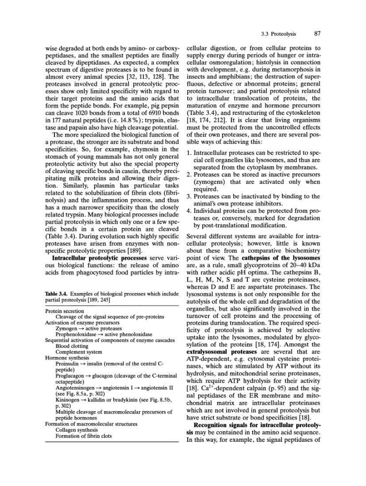
acids from phagocytosed food particles by intraTable 3.4. Examples of biological processes which include
partial proteolysis [189, 245]
Protein secretion
Cleavage of the signal sequence of pre-proteins
Activation of enzyme precursors
Zymogen ~ active proteases
Prophenoloxidase ~ active phenoloxidase
Sequential activation of components of enzyme cascades
Blood clotting
Complement system
Hormone synthesis
Proinsulin ~ insulin (removal of the central Cpeptide)
Proglucagon ~ glucagon (cleavage of the C-terminal
octapeptide)
Angiotensinogen ~ angiotensin I ~ angiotensin II
(see Fig. 8.Sa, p. 302)
Kininogen ~ kallidin or bradykinin (see Fig. 8.Sb,
p.302)
Multiple cleavage of macromolecular precursors of
peptide hormones
Formation of macromolecular structures
Collagen synthesis
Formation of fibrin clots
3.3 Proteolysis
87
cellular digestion, or from cellular proteins to
supply energy during periods of hunger or intracellular osmoregulation; histolysis in connection
with development, e.g. during metamorphosis in
insects and amphibians; the destruction of superfluous, defective or abnormal proteins; general
protein turnover; and partial proteolysis related
to intracellular translocation of proteins, the
maturation of enzyme and hormone precursors
(Table 3.4), and restructuring of the cytoskeleton
[18, 174, 212]. It is clear that living organisms
must be protected from the uncontrolled effects
of their own proteases, and there are several possible ways of achieving this:
1. Intracellular proteases can be restricted to special cell organelles like lysosomes, and thus are
separated from the cytoplasm by membranes.
2. Proteases can be stored as inactive precursors
(zymogens) that are activated only when
required.
3. Proteases can be inactivated by binding to the
animal's own protease inhibitors.
4. Individual proteins can be protected from proteases or, conversely, marked for degradation
by post-translational modification.
Several different systems are available for intracellular proteolysis; however, little is known
about these from a comparative biochemistry
point of view. The cathepsins of the Iysosomes
are, as a rule, small glycoproteins of 20-40 kDa
with rather acidic pH optima. The cathepsins B,
L, H, M, N, Sand T are cysteine proteinases,
whereas D and E are aspartate proteinases. The
lysosomal systems is not only responsible for the
autolysis of the whole cell and degradation of the
organelles, but also significantly involved in the
turnover of cell proteins and the processing of
proteins during translocation. The required specificity of proteolysis is achieved by selective
uptake into the lysosomes, modulated by glycosylation of the proteins [18, 174]. Amongst the
extralysosomal proteases are several that are
ATP-dependent, e.g. cytosomal cysteine proteinases, which are stimulated by ATP without its
hydrolysis, and mitochondrial serine proteinases,
which require ATP hydrolysis for their activity
[18]. Ca2+-dependent calpain (p.95) and the signal peptidases of the ER membrane and mitochondrial matrix are intracellular proteinases
which are not involved in general proteolysis but
have strict substrate or bond specificities [18].
Recognition signals for intracellular proteolysis may be contained in the amino acid sequence.
In this way, for example, the signal peptidases of
cleaved by dipeptidases. As expected, a complex
spectrum of digestive proteases is to be found in
almost every animal species [32, 113, 128]. The
proteases involved in general proteolytic processes show only limited specificity with regard to
their target proteins and the amino acids that
form the peptide bonds. For example, pig pepsin
can cleave 1020 bonds from a total of 6910 bonds
in 177 natural peptides (i.e. 14.8 %); trypsin, elastase and papain also have high cleavage potential.
The more specialized the biological function of
a protease, the stronger are its substrate and bond
specificities. So, for example, chymosin in the
stomach of young mammals has not only general
proteolytic activity but also the special property
of cleaving specific bonds in casein, thereby precipitating milk proteins and allowing their digestion. Similarly, plasmin has particular tasks
related to the solubilization of fibrin clots (fibrinolysis) and the inflammation process, and thus
has a much narrower specificity than the closely
related trypsin. Many biological processes include
partial proteolysis in which only one or a few specific bonds in a certain protein are cleaved
(Table 3.4). During evolution such highly specific
proteases have arisen from enzymes with nonspecific proteolytic properties [189].
Intracellular proteolytic processes serve various biological functions: the release of amino
acids from phagocytosed food particles by intraTable 3.4. Examples of biological processes which include
partial proteolysis [189, 245]
Protein secretion
Cleavage of the signal sequence of pre-proteins
Activation of enzyme precursors
Zymogen ~ active proteases
Prophenoloxidase ~ active phenoloxidase
Sequential activation of components of enzyme cascades
Blood clotting
Complement system
Hormone synthesis
Proinsulin ~ insulin (removal of the central Cpeptide)
Proglucagon ~ glucagon (cleavage of the C-terminal
octapeptide)
Angiotensinogen ~ angiotensin I ~ angiotensin II
(see Fig. 8.Sa, p. 302)
Kininogen ~ kallidin or bradykinin (see Fig. 8.Sb,
p.302)
Multiple cleavage of macromolecular precursors of
peptide hormones
Formation of macromolecular structures
Collagen synthesis
Formation of fibrin clots
3.3 Proteolysis
87
cellular digestion, or from cellular proteins to
supply energy during periods of hunger or intracellular osmoregulation; histolysis in connection
with development, e.g. during metamorphosis in
insects and amphibians; the destruction of superfluous, defective or abnormal proteins; general
protein turnover; and partial proteolysis related
to intracellular translocation of proteins, the
maturation of enzyme and hormone precursors
(Table 3.4), and restructuring of the cytoskeleton
[18, 174, 212]. It is clear that living organisms
must be protected from the uncontrolled effects
of their own proteases, and there are several possible ways of achieving this:
1. Intracellular proteases can be restricted to special cell organelles like lysosomes, and thus are
separated from the cytoplasm by membranes.
2. Proteases can be stored as inactive precursors
(zymogens) that are activated only when
required.
3. Proteases can be inactivated by binding to the
animal's own protease inhibitors.
4. Individual proteins can be protected from proteases or, conversely, marked for degradation
by post-translational modification.
Several different systems are available for intracellular proteolysis; however, little is known
about these from a comparative biochemistry
point of view. The cathepsins of the Iysosomes
are, as a rule, small glycoproteins of 20-40 kDa
with rather acidic pH optima. The cathepsins B,
L, H, M, N, Sand T are cysteine proteinases,
whereas D and E are aspartate proteinases. The
lysosomal systems is not only responsible for the
autolysis of the whole cell and degradation of the
organelles, but also significantly involved in the
turnover of cell proteins and the processing of
proteins during translocation. The required specificity of proteolysis is achieved by selective
uptake into the lysosomes, modulated by glycosylation of the proteins [18, 174]. Amongst the
extralysosomal proteases are several that are
ATP-dependent, e.g. cytosomal cysteine proteinases, which are stimulated by ATP without its
hydrolysis, and mitochondrial serine proteinases,
which require ATP hydrolysis for their activity
[18]. Ca2+-dependent calpain (p.95) and the signal peptidases of the ER membrane and mitochondrial matrix are intracellular proteinases
which are not involved in general proteolysis but
have strict substrate or bond specificities [18].
Recognition signals for intracellular proteolysis may be contained in the amino acid sequence.
In this way, for example, the signal peptidases of