

Chapter 3 Seagrass Morphology, Anatomy, and Ultrastructure
67
A
B
D
C
E
F
G
H
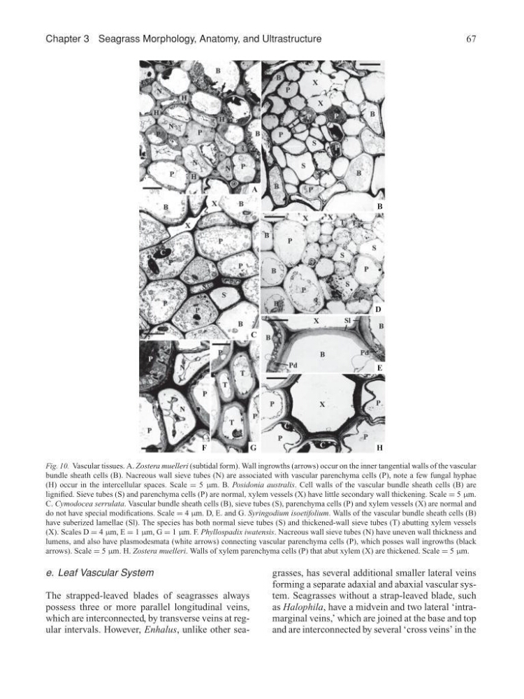
Fig. 10. Vascular tissues. A. Zostera muelleri (subtidal form). Wall ingrowths (arrows) occur on the inner tangential walls of the vascular
bundle sheath cells (B). Nacreous wall sieve tubes (N) are associated with vascular parenchyma cells (P), note a few fungal hyphae
(H) occur in the intercellular spaces. Scale = 5 µm. B. Posidonia australis. Cell walls of the vascular bundle sheath cells (B) are
lignified. Sieve tubes (S) and parenchyma cells (P) are normal, xylem vessels (X) have little secondary wall thickening. Scale = 5 µm.
C. Cymodocea serrulata. Vascular bundle sheath cells (B), sieve tubes (S), parenchyma cells (P) and xylem vessels (X) are normal and
do not have special modifications. Scale = 4 µm. D, E. and G. Syringodium isoetifolium. Walls of the vascular bundle sheath cells (B)
have suberized lamellae (Sl). The species has both normal sieve tubes (S) and thickened-wall sieve tubes (T) abutting xylem vessels
(X). Scales D = 4 µm, E = 1 µm, G = 1 µm. F. Phyllospadix iwatensis. Nacreous wall sieve tubes (N) have uneven wall thickness and
lumens, and also have plasmodesmata (white arrows) connecting vascular parenchyma cells (P), which posses wall ingrowths (black
arrows). Scale = 5 µm. H. Zostera muelleri. Walls of xylem parenchyma cells (P) that abut xylem (X) are thickened. Scale = 5 µm.
e. Leaf Vascular System
The strapped-leaved blades of seagrasses always
possess three or more parallel longitudinal veins,
which are interconnected, by transverse veins at regular intervals. However, Enhalus, unlike other seagrasses, has several additional smaller lateral veins
forming a separate adaxial and abaxial vascular system. Seagrasses without a strap-leaved blade, such
as Halophila, have a midvein and two lateral ‘intramarginal veins,’ which are joined at the base and top
and are interconnected by several ‘cross veins’ in the
67
A
B
D
C
E
F
G
H
Fig. 10. Vascular tissues. A. Zostera muelleri (subtidal form). Wall ingrowths (arrows) occur on the inner tangential walls of the vascular
bundle sheath cells (B). Nacreous wall sieve tubes (N) are associated with vascular parenchyma cells (P), note a few fungal hyphae
(H) occur in the intercellular spaces. Scale = 5 µm. B. Posidonia australis. Cell walls of the vascular bundle sheath cells (B) are
lignified. Sieve tubes (S) and parenchyma cells (P) are normal, xylem vessels (X) have little secondary wall thickening. Scale = 5 µm.
C. Cymodocea serrulata. Vascular bundle sheath cells (B), sieve tubes (S), parenchyma cells (P) and xylem vessels (X) are normal and
do not have special modifications. Scale = 4 µm. D, E. and G. Syringodium isoetifolium. Walls of the vascular bundle sheath cells (B)
have suberized lamellae (Sl). The species has both normal sieve tubes (S) and thickened-wall sieve tubes (T) abutting xylem vessels
(X). Scales D = 4 µm, E = 1 µm, G = 1 µm. F. Phyllospadix iwatensis. Nacreous wall sieve tubes (N) have uneven wall thickness and
lumens, and also have plasmodesmata (white arrows) connecting vascular parenchyma cells (P), which posses wall ingrowths (black
arrows). Scale = 5 µm. H. Zostera muelleri. Walls of xylem parenchyma cells (P) that abut xylem (X) are thickened. Scale = 5 µm.
e. Leaf Vascular System
The strapped-leaved blades of seagrasses always
possess three or more parallel longitudinal veins,
which are interconnected, by transverse veins at regular intervals. However, Enhalus, unlike other seagrasses, has several additional smaller lateral veins
forming a separate adaxial and abaxial vascular system. Seagrasses without a strap-leaved blade, such
as Halophila, have a midvein and two lateral ‘intramarginal veins,’ which are joined at the base and top
and are interconnected by several ‘cross veins’ in the