

Chapter 3 Seagrass Morphology, Anatomy, and Ultrastructure
65
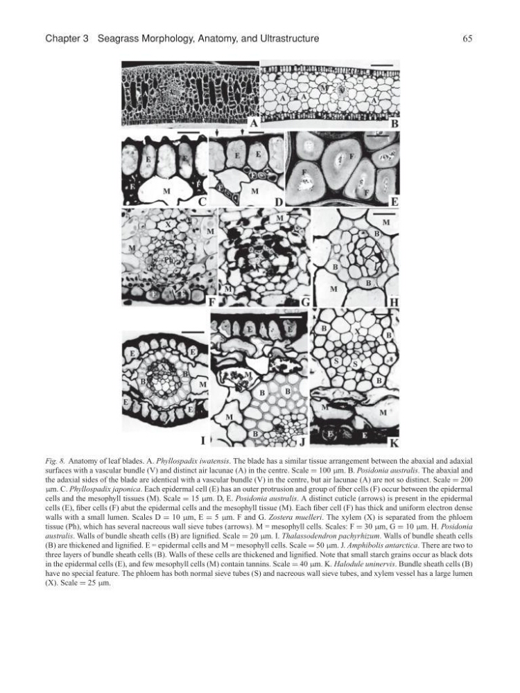
Fig. 8. Anatomy of leaf blades. A. Phyllospadix iwatensis. The blade has a similar tissue arrangement between the abaxial and adaxial
surfaces with a vascular bundle (V) and distinct air lacunae (A) in the centre. Scale = 100 µm. B. Posidonia australis. The abaxial and
the adaxial sides of the blade are identical with a vascular bundle (V) in the centre, but air lacunae (A) are not so distinct. Scale = 200
µm. C. Phyllospadix japonica. Each epidermal cell (E) has an outer protrusion and group of fiber cells (F) occur between the epidermal
cells and the mesophyll tissues (M). Scale = 15 µm. D, E. Posidonia australis. A distinct cuticle (arrows) is present in the epidermal
cells (E), fiber cells (F) abut the epidermal cells and the mesophyll tissue (M). Each fiber cell (F) has thick and uniform electron dense
walls with a small lumen. Scales D = 10 µm, E = 5 µm. F and G. Zostera muelleri. The xylem (X) is separated from the phloem
tissue (Ph), which has several nacreous wall sieve tubes (arrows). M = mesophyll cells. Scales: F = 30 µm, G = 10 µm. H. Posidonia
australis. Walls of bundle sheath cells (B) are lignified. Scale = 20 µm. I. Thalassodendron pachyrhizum. Walls of bundle sheath cells
(B) are thickened and lignified. E = epidermal cells and M = mesophyll cells. Scale = 50 µm. J. Amphibolis antarctica. There are two to
three layers of bundle sheath cells (B). Walls of these cells are thickened and lignified. Note that small starch grains occur as black dots
in the epidermal cells (E), and few mesophyll cells (M) contain tannins. Scale = 40 µm. K. Halodule uninervis. Bundle sheath cells (B)
have no special feature. The phloem has both normal sieve tubes (S) and nacreous wall sieve tubes, and xylem vessel has a large lumen
(X). Scale = 25 µm.
65
Fig. 8. Anatomy of leaf blades. A. Phyllospadix iwatensis. The blade has a similar tissue arrangement between the abaxial and adaxial
surfaces with a vascular bundle (V) and distinct air lacunae (A) in the centre. Scale = 100 µm. B. Posidonia australis. The abaxial and
the adaxial sides of the blade are identical with a vascular bundle (V) in the centre, but air lacunae (A) are not so distinct. Scale = 200
µm. C. Phyllospadix japonica. Each epidermal cell (E) has an outer protrusion and group of fiber cells (F) occur between the epidermal
cells and the mesophyll tissues (M). Scale = 15 µm. D, E. Posidonia australis. A distinct cuticle (arrows) is present in the epidermal
cells (E), fiber cells (F) abut the epidermal cells and the mesophyll tissue (M). Each fiber cell (F) has thick and uniform electron dense
walls with a small lumen. Scales D = 10 µm, E = 5 µm. F and G. Zostera muelleri. The xylem (X) is separated from the phloem
tissue (Ph), which has several nacreous wall sieve tubes (arrows). M = mesophyll cells. Scales: F = 30 µm, G = 10 µm. H. Posidonia
australis. Walls of bundle sheath cells (B) are lignified. Scale = 20 µm. I. Thalassodendron pachyrhizum. Walls of bundle sheath cells
(B) are thickened and lignified. E = epidermal cells and M = mesophyll cells. Scale = 50 µm. J. Amphibolis antarctica. There are two to
three layers of bundle sheath cells (B). Walls of these cells are thickened and lignified. Note that small starch grains occur as black dots
in the epidermal cells (E), and few mesophyll cells (M) contain tannins. Scale = 40 µm. K. Halodule uninervis. Bundle sheath cells (B)
have no special feature. The phloem has both normal sieve tubes (S) and nacreous wall sieve tubes, and xylem vessel has a large lumen
(X). Scale = 25 µm.