

H 2 , organic compounds). Anoxygenic phototrophic bacteria
so have an anaerobic photosynthetic activity.
Anoxygenic phototrophic activity (photosystem of type II)
was demonstrated in planktonic aerobic heterotrophic bacteria
or aerobic anoxygenic phototrophs (Erythrobacter,
Roseobacter, Erythromicrobium, Roseococcus, Porphyrobacter, etc.). However, photophosphorylations only
provide a supplementary supply of energy, unable to ensure
by itself the growth of these microorganisms (Yurkov and
Beatty 1998; Fuchs et al. 2007).
The BChl a (P870 or P840) excited by light transfers its
electrons to an electron acceptor at low potential (Bph or
BChl 663). The electrons are then transferred to a series of
electron carriers. During this transfer, the electron energy
level drops. Finally, these low-energy electrons return to the
reaction centers and reduce again the Bchl. During the cycle,
protons are transferred to the outside of the cytoplasmic
membrane. H
+ gradient is thus created; it is at the origin of
a proton-motive force that generates ATP by photophosphorylation. The reduction of NAD
+ is carried by electrons
coming out of cyclical flow, thus causing a deficit of
electrons. This is compensated by a contribution from inorganic external donors (reduced sulfur compounds, H 2 ) or
organic (succinate, malate) via the cytochromes C555 or
C2 in the case of sulfur compounds and plastoquinone
(PQ) in that of the succinate. The electrons return to reaction
centers where they make up the deficit of BChl which is
consecutive to its excitement. In the case of phototrophic
purple bacteria, the electrons which reduce coenzymes pass
via the complex cytochrome b/c1 (Fig. 3.29a). The positive
redox potential of this complex makes impossible the direct
reduction of NAD
+ whose redox potential is very negative
(À0.32 V). A reverse flow of electrons that consumes energy
is needed to allow the reduction of coenzymes. In
phototrophic green bacteria, the redox potential of ferredoxin is sufficiently electronegative (À0.42 V) to directly
reduce coenzymes (Fig. 3.29b).
3.3.4.4 Use of Light Energy by the Archaea
Some extreme halophilic archaea, Halobacterium salinarum
in particular, can use light as an energy source when dioxygen
levels in their natural environment are too low. The membrane of archaea is largely invaded by dark red color that
characterizes the purple membrane containing a colored
protein, bacteriorhodopsin, near the rhodopsin of the retina
of eyes. Bacteriorhodopsin has a carotenoid as prosthetic
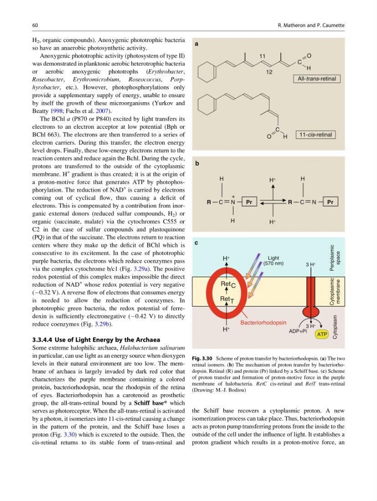
group, the all-trans-retinal bound by a Schiff base* which
serves as photoreceptor. When the all-trans-retinal is activated
by a photon, it isomerizes into 11-cis-retinal causing a change
in the pattern of the protein, and the Schiff base loses a
proton (Fig. 3.30) which is excreted to the outside. Then, the
cis-retinal returns to its stable form of trans-retinal and
the Schiff base recovers a cytoplasmic proton. A new
isomerization process can take place. Thus, bacteriorhodopsin
acts as proton pump transferring protons from the inside to the
outside of the cell under the influence of light. It establishes a
proton gradient which results in a proton-motive force, an
11
12
O
H
O
H
11-cis-retinal
C
C
All-trans-retinal
a
R
H
H
+
C N
Pr
+
H
+
H
R
H
C N
Pr
b
H
+
H
+
c
ADP+Pi
3 H
+
3 H
+
Ret
Ret
C
T
Periplasmic
space
Cytoplasmic
membrane
Cytoplasm
Bacteriorhodopsin
Light
(570 nm)
ATP
Fig. 3.30 Scheme of proton transfer by bacteriorhodopsin. (a) The two
retinal isomers. (b) The mechanism of proton transfer by bacteriorhodopsin. Retinal (R) and protein (Pr) linked by a Schiff base. (c) Scheme
of proton transfer and formation of proton-motive force in the purple
membrane of halobacteria. RetC cis-retinal and RetT trans-retinal
(Drawing: M.-J. Bodiou)
60
R. Matheron and P. Caumette
so have an anaerobic photosynthetic activity.
Anoxygenic phototrophic activity (photosystem of type II)
was demonstrated in planktonic aerobic heterotrophic bacteria
or aerobic anoxygenic phototrophs (Erythrobacter,
Roseobacter, Erythromicrobium, Roseococcus, Porphyrobacter, etc.). However, photophosphorylations only
provide a supplementary supply of energy, unable to ensure
by itself the growth of these microorganisms (Yurkov and
Beatty 1998; Fuchs et al. 2007).
The BChl a (P870 or P840) excited by light transfers its
electrons to an electron acceptor at low potential (Bph or
BChl 663). The electrons are then transferred to a series of
electron carriers. During this transfer, the electron energy
level drops. Finally, these low-energy electrons return to the
reaction centers and reduce again the Bchl. During the cycle,
protons are transferred to the outside of the cytoplasmic
membrane. H
+ gradient is thus created; it is at the origin of
a proton-motive force that generates ATP by photophosphorylation. The reduction of NAD
+ is carried by electrons
coming out of cyclical flow, thus causing a deficit of
electrons. This is compensated by a contribution from inorganic external donors (reduced sulfur compounds, H 2 ) or
organic (succinate, malate) via the cytochromes C555 or
C2 in the case of sulfur compounds and plastoquinone
(PQ) in that of the succinate. The electrons return to reaction
centers where they make up the deficit of BChl which is
consecutive to its excitement. In the case of phototrophic
purple bacteria, the electrons which reduce coenzymes pass
via the complex cytochrome b/c1 (Fig. 3.29a). The positive
redox potential of this complex makes impossible the direct
reduction of NAD
+ whose redox potential is very negative
(À0.32 V). A reverse flow of electrons that consumes energy
is needed to allow the reduction of coenzymes. In
phototrophic green bacteria, the redox potential of ferredoxin is sufficiently electronegative (À0.42 V) to directly
reduce coenzymes (Fig. 3.29b).
3.3.4.4 Use of Light Energy by the Archaea
Some extreme halophilic archaea, Halobacterium salinarum
in particular, can use light as an energy source when dioxygen
levels in their natural environment are too low. The membrane of archaea is largely invaded by dark red color that
characterizes the purple membrane containing a colored
protein, bacteriorhodopsin, near the rhodopsin of the retina
of eyes. Bacteriorhodopsin has a carotenoid as prosthetic
group, the all-trans-retinal bound by a Schiff base* which
serves as photoreceptor. When the all-trans-retinal is activated
by a photon, it isomerizes into 11-cis-retinal causing a change
in the pattern of the protein, and the Schiff base loses a
proton (Fig. 3.30) which is excreted to the outside. Then, the
cis-retinal returns to its stable form of trans-retinal and
the Schiff base recovers a cytoplasmic proton. A new
isomerization process can take place. Thus, bacteriorhodopsin
acts as proton pump transferring protons from the inside to the
outside of the cell under the influence of light. It establishes a
proton gradient which results in a proton-motive force, an
11
12
O
H
O
H
11-cis-retinal
C
C
All-trans-retinal
a
R
H
H
+
C N
Pr
+
H
+
H
R
H
C N
Pr
b
H
+
H
+
c
ADP+Pi
3 H
+
3 H
+
Ret
Ret
C
T
Periplasmic
space
Cytoplasmic
membrane
Cytoplasm
Bacteriorhodopsin
Light
(570 nm)
ATP
Fig. 3.30 Scheme of proton transfer by bacteriorhodopsin. (a) The two
retinal isomers. (b) The mechanism of proton transfer by bacteriorhodopsin. Retinal (R) and protein (Pr) linked by a Schiff base. (c) Scheme
of proton transfer and formation of proton-motive force in the purple
membrane of halobacteria. RetC cis-retinal and RetT trans-retinal
(Drawing: M.-J. Bodiou)
60
R. Matheron and P. Caumette