

14.4.4.3 Photosynthetic Microbial Mats
Microbial mats are widely scattered across the globe and can
be considered as actual stromatolites, the oldest of which
dating from 3.5 billion years (cf. Sect. 4.2.2). The ubiquity
and the presence of these ecosystems for several millions of
years exhibit their capability to adapt to different
environments, including hostile and fluctuating conditions.
They can indeed be observed in a wide range of
environments, including coastal, hypersaline, polar lake
and soil, hot spring, or hypersaline lake environments.
These organo-sedimentary structures have a role in
stabilizing coastal areas subjected to erosion by waves,
tides, and winds.
Microbial mats are perfectly organized ecosystems in
which the interactions between different functional groups
are very strong (cf. Sect. 9.7.3). They constitute an excellent
example of microbial consortium which combines different
populations and is organized in the form of horizontal strata,
due to physicochemical gradients of light, dioxygen, and sulfur (Fig. 14.44). In microbial mats, microorganisms of simple
structure (compared with multicellular organisms) are
organized in a form of ectosymbiosis* made by genetically
Box 14.13 (continued)
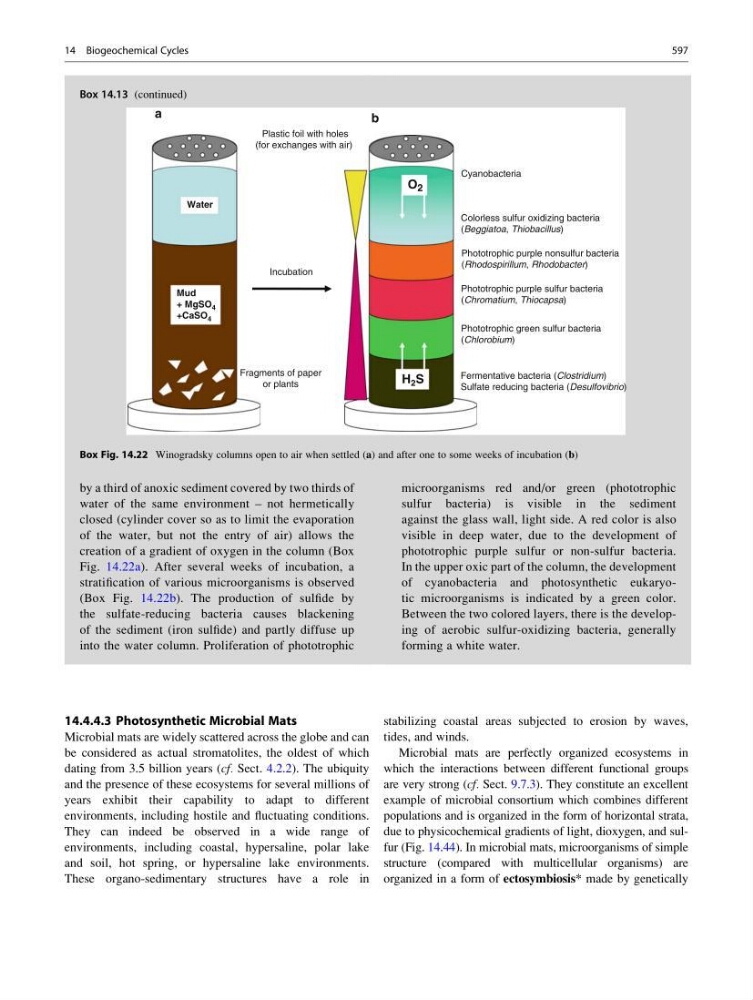
by a third of anoxic sediment covered by two thirds of
water of the same environment – not hermetically
closed (cylinder cover so as to limit the evaporation
of the water, but not the entry of air) allows the
creation of a gradient of oxygen in the column (Box
Fig. 14.22a). After several weeks of incubation, a
stratification of various microorganisms is observed
(Box Fig. 14.22b). The production of sulfide by
the sulfate-reducing bacteria causes blackening
of the sediment (iron sulfide) and partly diffuse up
into the water column. Proliferation of phototrophic
(continued)
Box 14.13 (continued)
microorganisms red and/or green (phototrophic
sulfur bacteria) is visible in the sediment
against the glass wall, light side. A red color is also
visible in deep water, due to the development of
phototrophic purple sulfur or non-sulfur bacteria.
In the upper oxic part of the column, the development
of cyanobacteria and photosynthetic eukaryotic microorganisms is indicated by a green color.
Between the two colored layers, there is the developing of aerobic sulfur-oxidizing bacteria, generally
forming a white water.
Incubation
Fragments of paper
or plants
Mud
+ MgSO 4
+CaSO 4
Water
Fermentative bacteria (Clostridium)
Sulfate reducing bacteria (Desulfovibrio)
Phototrophic green sulfur bacteria
(Chlorobium)
Phototrophic purple sulfur bacteria
(Chromatium, Thiocapsa)
Phototrophic purple nonsulfur bacteria
(Rhodospirillum, Rhodobacter)
Colorless sulfur oxidizing bacteria
(Beggiatoa, Thiobacillus)
Cyanobacteria
H 2 S
a
b
Plastic foil with holes
(for exchanges with air)
O 2
Box Fig. 14.22 Winogradsky columns open to air when settled (a) and after one to some weeks of incubation (b)
14 Biogeochemical Cycles
597