

especially in intracellular microbes (Roulston et al. 1999;
Gao and Abu Kwaid 2000).
Inhibition of Apoptosis by Microorganisms
Commensal and pathogenic bacteria can inactivate apoptosis
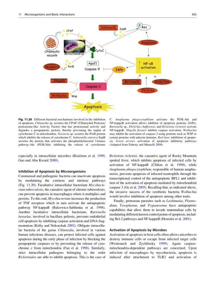
by modulating the extrinsic and intrinsic pathways
(Fig. 11.20). Facultative intracellular bacterium Mycobacterium tuberculosis, the causative agent of chronic tuberculosis,
can prevent apoptosis in macrophages where it multiplies and
persists. To this end, Mycobacterium increases the production
of TNF receptors which in turn activate the antiapoptotic
pathway NF-kappaB (Balcewicz-Sablinska et al. 1998).
Another facultative intracellular bacterium, Bartonella
henselae, involved in bacillary peliosis, prevents endothelial
cell apoptosis by inhibiting caspase activation and DNA fragmentation (Kirby and Nekorchuk 2002). Obligate intracellular bacteria of the genus Chlamydia, involved in various
human infectious diseases, can protect infected cells against
apoptosis during the early phase of infection by blocking the
proapoptotic caspases or by preventing the release of cytochrome c from mitochondria (Fan et al. 1998). Similarly,
strict intracellular pathogens belonging to the order
Rickettsiales are able to inhibit apoptosis. This is the case of
Rickettsia rickettsii, the causative agent of Rocky Mountain
spotted fever, which inhibits apoptosis of infected cells by
activation of NF-kappaB (Clifton et al. 1998), while
Anaplasma phagocytophilum, responsible of human anaplasmosis, prevents apoptosis of infected neutrophils through the
transcriptional control of the antiapoptotic BFL1 and inhibition of the activation of apoptosis mediated by mitochondrial
caspase 3 (Ge et al. 2005). Recalling that, as indicated above,
the invasive success of the symbiotic bacteria Wolbachia
would involve inhibition of apoptosis among other traits.
Finally, protozoan parasites such as Leishmania, Plasmodium, Toxoplasma, and Trypanosoma have antiapoptotic
capabilities that allow them to invade mammalian cells by
modulating different known control points of apoptosis, including Bcl-2 pathways and NF-kappaB (Heussler et al. 2001).
Activation of Apoptosis by Microbes
Activation of apoptosis in host cells often allows microbes to
destroy immune cells or escape from infected target cells
(Weinrauch and Zychlinsky 1999). Again caspasemitochondria-dependent pathways are concerned. Upon
infection of macrophages by mycobacteria, apoptosis is
induced after attachment to TLR2 and activation of
Fig. 11.20 Different bacterial mechanisms involved in the inhibition
of apoptosis. Chlamydia sp. secretes the CPAF (Chlamydial Protease/
proteasome-like Activity Factor) that has proteasomal activity and
degrades a proapoptotic protein, thereby preventing the output of
cytochrome C in mitochondria. Neisseria sp. secretes the PorB protein
which inhibits the release of cytochrome C. Salmonella enterica SopB
secretes the protein that activates the phosphatidylinositol 3-kinase
pathway-Akt (PI3K/Akt) inhibiting the release of cytochrome
C. Anaplasma phagocytophilum activates the PI3K-Akt and
NF-kappaB activation allows inhibitor of apoptosis proteins (IAPs).
Bartonella sp., Ehrlichia chaffeensis, and Rickettsia rickettsii activate
NF-kappaB. Shigella flexneri inhibits caspase activation. Wolbachia
may inhibit the activation of caspase-3 using proteins such as WSP or
certain proteins with ankyrin domains. Red lines: inhibition of apoptosis. Green arrows: activation of apoptosis inhibitory pathways
(Adapted from Faherty and Maurelli 2008)
11 Microorganisms and Biotic Interactions
435
Gao and Abu Kwaid 2000).
Inhibition of Apoptosis by Microorganisms
Commensal and pathogenic bacteria can inactivate apoptosis
by modulating the extrinsic and intrinsic pathways
(Fig. 11.20). Facultative intracellular bacterium Mycobacterium tuberculosis, the causative agent of chronic tuberculosis,
can prevent apoptosis in macrophages where it multiplies and
persists. To this end, Mycobacterium increases the production
of TNF receptors which in turn activate the antiapoptotic
pathway NF-kappaB (Balcewicz-Sablinska et al. 1998).
Another facultative intracellular bacterium, Bartonella
henselae, involved in bacillary peliosis, prevents endothelial
cell apoptosis by inhibiting caspase activation and DNA fragmentation (Kirby and Nekorchuk 2002). Obligate intracellular bacteria of the genus Chlamydia, involved in various
human infectious diseases, can protect infected cells against
apoptosis during the early phase of infection by blocking the
proapoptotic caspases or by preventing the release of cytochrome c from mitochondria (Fan et al. 1998). Similarly,
strict intracellular pathogens belonging to the order
Rickettsiales are able to inhibit apoptosis. This is the case of
Rickettsia rickettsii, the causative agent of Rocky Mountain
spotted fever, which inhibits apoptosis of infected cells by
activation of NF-kappaB (Clifton et al. 1998), while
Anaplasma phagocytophilum, responsible of human anaplasmosis, prevents apoptosis of infected neutrophils through the
transcriptional control of the antiapoptotic BFL1 and inhibition of the activation of apoptosis mediated by mitochondrial
caspase 3 (Ge et al. 2005). Recalling that, as indicated above,
the invasive success of the symbiotic bacteria Wolbachia
would involve inhibition of apoptosis among other traits.
Finally, protozoan parasites such as Leishmania, Plasmodium, Toxoplasma, and Trypanosoma have antiapoptotic
capabilities that allow them to invade mammalian cells by
modulating different known control points of apoptosis, including Bcl-2 pathways and NF-kappaB (Heussler et al. 2001).
Activation of Apoptosis by Microbes
Activation of apoptosis in host cells often allows microbes to
destroy immune cells or escape from infected target cells
(Weinrauch and Zychlinsky 1999). Again caspasemitochondria-dependent pathways are concerned. Upon
infection of macrophages by mycobacteria, apoptosis is
induced after attachment to TLR2 and activation of
Fig. 11.20 Different bacterial mechanisms involved in the inhibition
of apoptosis. Chlamydia sp. secretes the CPAF (Chlamydial Protease/
proteasome-like Activity Factor) that has proteasomal activity and
degrades a proapoptotic protein, thereby preventing the output of
cytochrome C in mitochondria. Neisseria sp. secretes the PorB protein
which inhibits the release of cytochrome C. Salmonella enterica SopB
secretes the protein that activates the phosphatidylinositol 3-kinase
pathway-Akt (PI3K/Akt) inhibiting the release of cytochrome
C. Anaplasma phagocytophilum activates the PI3K-Akt and
NF-kappaB activation allows inhibitor of apoptosis proteins (IAPs).
Bartonella sp., Ehrlichia chaffeensis, and Rickettsia rickettsii activate
NF-kappaB. Shigella flexneri inhibits caspase activation. Wolbachia
may inhibit the activation of caspase-3 using proteins such as WSP or
certain proteins with ankyrin domains. Red lines: inhibition of apoptosis. Green arrows: activation of apoptosis inhibitory pathways
(Adapted from Faherty and Maurelli 2008)
11 Microorganisms and Biotic Interactions
435