

response to microbial infection (Salaun et al. 2007). For
example, the perception of bacterial LPS by TLR4 on endothelial cells leads to the induction of apoptosis (Bannerman
and Goldblum 1997; Aliprantis et al. 1999). In mammalian
cells such as mice, apoptosis mediated by TLR4 confers
resistance to infection due to the perception of the pneumococcal virulence factor, pneumolysin (Srivastava et al.
2005). To eliminate or stop the progression of the infection
with Shigella or Mycobacterium, infected macrophages can
undergo apoptosis resulting in concomitant death of these
bacterial pathogens (Zychlinsky and Sansonetti 1997).
In insects, the involvement of apoptosis in the defense
mechanism was shown for the first time in response to
infection of the butterfly Spodoptera frugiperda by baculovirus (Clarke and Clem 2003). Phenomena associated with
apoptotic caspase activity were observed in mosquitoes
infected with Plasmodium (Hurd et al. 2006).
11.4.4.2 Modulation of Apoptosis
by Microorganisms
Microbial pathogens have developed various mechanisms to
inhibit apoptosis in host cells to facilitate their replication
and persistence. Microbes can also induce apoptosis in
immune cells to destroy these cells thereby facilitating
their escape. The ability to circumvent host defenses by
modulating apoptosis is found in various microbial taxa,
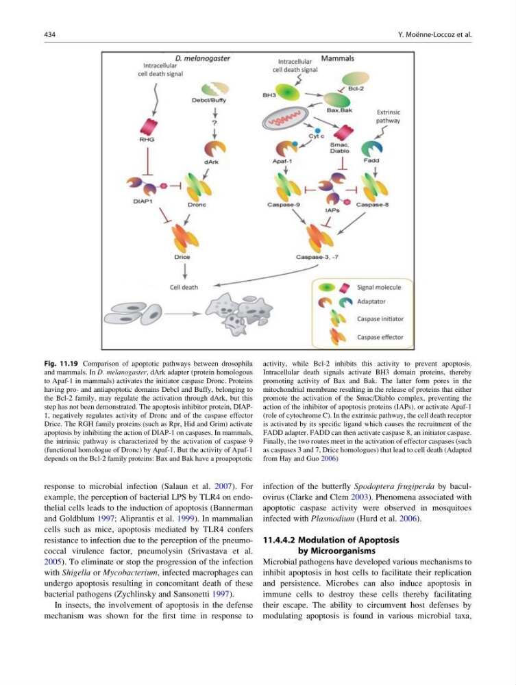
Fig. 11.19 Comparison of apoptotic pathways between drosophila
and mammals. In D. melanogaster, dArk adapter (protein homologous
to Apaf-1 in mammals) activates the initiator caspase Dronc. Proteins
having pro- and antiapoptotic domains Debcl and Buffy, belonging to
the Bcl-2 family, may regulate the activation through dArk, but this
step has not been demonstrated. The apoptosis inhibitor protein, DIAP1, negatively regulates activity of Dronc and of the caspase effector
Drice. The RGH family proteins (such as Rpr, Hid and Grim) activate
apoptosis by inhibiting the action of DIAP-1 on caspases. In mammals,
the intrinsic pathway is characterized by the activation of caspase 9
(functional homologue of Dronc) by Apaf-1. But the activity of Apaf-1
depends on the Bcl-2 family proteins: Bax and Bak have a proapoptotic
activity, while Bcl-2 inhibits this activity to prevent apoptosis.
Intracellular death signals activate BH3 domain proteins, thereby
promoting activity of Bax and Bak. The latter form pores in the
mitochondrial membrane resulting in the release of proteins that either
promote the activation of the Smac/Diablo complex, preventing the
action of the inhibitor of apoptosis proteins (IAPs), or activate Apaf-1
(role of cytochrome C). In the extrinsic pathway, the cell death receptor
is activated by its specific ligand which causes the recruitment of the
FADD adapter. FADD can then activate caspase 8, an initiator caspase.
Finally, the two routes meet in the activation of effector caspases (such
as caspases 3 and 7, Drice homologues) that lead to cell death (Adapted
from Hay and Guo 2006)
434
Y. Moe ¨nne-Loccoz et al.
example, the perception of bacterial LPS by TLR4 on endothelial cells leads to the induction of apoptosis (Bannerman
and Goldblum 1997; Aliprantis et al. 1999). In mammalian
cells such as mice, apoptosis mediated by TLR4 confers
resistance to infection due to the perception of the pneumococcal virulence factor, pneumolysin (Srivastava et al.
2005). To eliminate or stop the progression of the infection
with Shigella or Mycobacterium, infected macrophages can
undergo apoptosis resulting in concomitant death of these
bacterial pathogens (Zychlinsky and Sansonetti 1997).
In insects, the involvement of apoptosis in the defense
mechanism was shown for the first time in response to
infection of the butterfly Spodoptera frugiperda by baculovirus (Clarke and Clem 2003). Phenomena associated with
apoptotic caspase activity were observed in mosquitoes
infected with Plasmodium (Hurd et al. 2006).
11.4.4.2 Modulation of Apoptosis
by Microorganisms
Microbial pathogens have developed various mechanisms to
inhibit apoptosis in host cells to facilitate their replication
and persistence. Microbes can also induce apoptosis in
immune cells to destroy these cells thereby facilitating
their escape. The ability to circumvent host defenses by
modulating apoptosis is found in various microbial taxa,
Fig. 11.19 Comparison of apoptotic pathways between drosophila
and mammals. In D. melanogaster, dArk adapter (protein homologous
to Apaf-1 in mammals) activates the initiator caspase Dronc. Proteins
having pro- and antiapoptotic domains Debcl and Buffy, belonging to
the Bcl-2 family, may regulate the activation through dArk, but this
step has not been demonstrated. The apoptosis inhibitor protein, DIAP1, negatively regulates activity of Dronc and of the caspase effector
Drice. The RGH family proteins (such as Rpr, Hid and Grim) activate
apoptosis by inhibiting the action of DIAP-1 on caspases. In mammals,
the intrinsic pathway is characterized by the activation of caspase 9
(functional homologue of Dronc) by Apaf-1. But the activity of Apaf-1
depends on the Bcl-2 family proteins: Bax and Bak have a proapoptotic
activity, while Bcl-2 inhibits this activity to prevent apoptosis.
Intracellular death signals activate BH3 domain proteins, thereby
promoting activity of Bax and Bak. The latter form pores in the
mitochondrial membrane resulting in the release of proteins that either
promote the activation of the Smac/Diablo complex, preventing the
action of the inhibitor of apoptosis proteins (IAPs), or activate Apaf-1
(role of cytochrome C). In the extrinsic pathway, the cell death receptor
is activated by its specific ligand which causes the recruitment of the
FADD adapter. FADD can then activate caspase 8, an initiator caspase.
Finally, the two routes meet in the activation of effector caspases (such
as caspases 3 and 7, Drice homologues) that lead to cell death (Adapted
from Hay and Guo 2006)
434
Y. Moe ¨nne-Loccoz et al.