

of cells, some with functional specificity, the first step toward
multicellularity (cf. Sect. 5.2). The cells of prokaryotes are
generally small, close to the micrometer, spherical (cocci) or
elongated rod shaped, straight, curved, or spiral. Particular
forms have stalks or protuberances (Fig. 3.2). Some prokaryotic cells may have large sizes, visible with the naked eye
(such as Thiomargarita namibiensis, 300–750 μm in diameter)
(cf. Chap. 5, Fig. 5.1c); in contrast, nanobacteria whose cells
are less than 0.2 μm in diameter were isolated from different
environments.
Exchanges between cells and the external environment
are realized at the cell surface. The surface/volume ratio
which is inversely proportional to cell size gives an advantage to prokaryotes compared to all other living organisms.
However, this feature makes it more sensitive to changes in
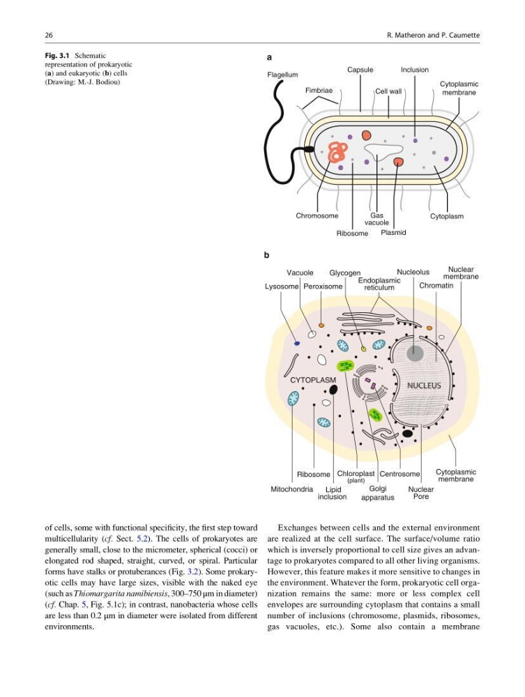
the environment. Whatever the form, prokaryotic cell organization remains the same: more or less complex cell
envelopes are surrounding cytoplasm that contains a small
number of inclusions (chromosome, plasmids, ribosomes,
gas vacuoles, etc.). Some also contain a membrane
.
.
.
.
.
.
.
.
.
Flagellum
Fimbriae
Capsule
Cell wall
Cytoplasmic
membrane
Inclusion
Ribosome
Plasmid
Cytoplasm
Gas
vacuole
Chromosome
. .
.
. .
.
a
. .
.
.
. . . .
.
.
.
.
.
.
.
.
.
.
.
.
.
.
.
.
.
.
.
.
.
.
. .
.
.
. .
.
.
.
. .
. . . . . .
. .
Centrosome
Ribosome
Mitochondria
Lipid
inclusion
Nuclear
membrane
Chromatin
Nucleolus
Endoplasmic
reticulum
Glycogen
Lysosome
Vacuole
Peroxisome
Chloroplast
Golgi
apparatus
Nuclear
Pore
Cytoplasmic
membrane
CYTOPLASM
NUCLEUS
(plant)
b
Fig. 3.1 Schematic
representation of prokaryotic
(a) and eukaryotic (b) cells
(Drawing: M.-J. Bodiou)
26
R. Matheron and P. Caumette
multicellularity (cf. Sect. 5.2). The cells of prokaryotes are
generally small, close to the micrometer, spherical (cocci) or
elongated rod shaped, straight, curved, or spiral. Particular
forms have stalks or protuberances (Fig. 3.2). Some prokaryotic cells may have large sizes, visible with the naked eye
(such as Thiomargarita namibiensis, 300–750 μm in diameter)
(cf. Chap. 5, Fig. 5.1c); in contrast, nanobacteria whose cells
are less than 0.2 μm in diameter were isolated from different
environments.
Exchanges between cells and the external environment
are realized at the cell surface. The surface/volume ratio
which is inversely proportional to cell size gives an advantage to prokaryotes compared to all other living organisms.
However, this feature makes it more sensitive to changes in
the environment. Whatever the form, prokaryotic cell organization remains the same: more or less complex cell
envelopes are surrounding cytoplasm that contains a small
number of inclusions (chromosome, plasmids, ribosomes,
gas vacuoles, etc.). Some also contain a membrane
.
.
.
.
.
.
.
.
.
Flagellum
Fimbriae
Capsule
Cell wall
Cytoplasmic
membrane
Inclusion
Ribosome
Plasmid
Cytoplasm
Gas
vacuole
Chromosome
. .
.
. .
.
a
. .
.
.
. . . .
.
.
.
.
.
.
.
.
.
.
.
.
.
.
.
.
.
.
.
.
.
.
. .
.
.
. .
.
.
.
. .
. . . . . .
. .
Centrosome
Ribosome
Mitochondria
Lipid
inclusion
Nuclear
membrane
Chromatin
Nucleolus
Endoplasmic
reticulum
Glycogen
Lysosome
Vacuole
Peroxisome
Chloroplast
Golgi
apparatus
Nuclear
Pore
Cytoplasmic
membrane
CYTOPLASM
NUCLEUS
(plant)
b
Fig. 3.1 Schematic
representation of prokaryotic
(a) and eukaryotic (b) cells
(Drawing: M.-J. Bodiou)
26
R. Matheron and P. Caumette