

teeth). One common example is the formation of dental
plaque, which begins with the formation of a biofilm by
pioneer bacteria or early colonizers (Streptococcus oralis,
Stomatococcus) that adhere to the teeth and produce the
biofilm matrix. Then, other bacteria (staphylococci, Enterobacteriaceae, fermentative bacteria) grow on the first
deposit (secondary colonizers) and cause, through their
development, the formation of anoxic zones in the deepest
part in contact with the tooth where fermentation produces
organic acids and releases sulfides that are responsible for
the attack of the tooth. Biofilms are also often installed on
inert surfaces. In tubes where liquids circulate, many types
of biofilms are common. Pseudomonas aeruginosa is
responsible for the formation of catheters biofilms in hospital settings, making it the top infectious agent responsible for
nosocomial infections. Heterotrophic bacteria are responsible for aerobic and anaerobic biofilms in water pipes, gas,
and oil and are often the cause of bacterial corrosion
problems in pipelines (sulfate-reducing bacteria, Box
14.12). Microbial biofilms also form on submerged
structures (metal or concrete) in ports, boats, and marine
platforms and are responsible for corrosion.
Microbial Mats
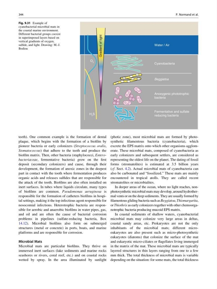
Microbial mats are particular biofilms. They thrive on
immersed inert surfaces (lake sediments and marine rocks
seashores or rivers, coral reef, etc.) and on coastal rocks
wetted by spray. In the area illuminated by sunlight
(photic zone), most microbial mats are formed by photosynthetic filamentous bacteria (cyanobacteria), which
excrete the EPS matrix onto which other organisms agglomerate. These microbial mats, composed of cyanobacteria as
early colonizers and subsequent settlers, are considered as
representing the oldest life on the planet. The dating of fossil
forms (stromatolites) is estimated at 3.5 billion years
(cf. Sect. 4.2). Actual microbial mats of cyanobacteria can
also be carbonated and “fossilized.” These mats are mainly
encountered in tropical atolls. They are called recent
stromatolites or microbialites.
In deeper areas of the ocean, where no light reaches, nonphotosynthetic microbial mats may develop, around hydrothermal vents or on the deep sediments. They are usually formed by
filamentous gliding bacteria such as Beggiatoa, Thiomargarita,
or Thiothrix as early colonizers together with other chemoorganotrophic bacteria producing mucoid EPS matrix.
In coastal sediments of shallow waters, cyanobacterial
microbial mats may colonize very large areas in deltas,
coastal sandy areas, etc. Prokaryotes are not the only
inhabitants of the microbial mats; different microeukaryotes are also present such as micro-photosynthetic
eukaryotes (diatoms) that colonize the surface of the mat
and eukaryotic micro-ciliates or flagellates living immerged
in the matrix of the mat. These microbial mats are typically
layered structures in thin layers ranging from one to a few
mm thick. The total thickness of microbial mats is variable
depending on the situation: for some mats, the total thickness
Water / Air
Cyanobacteria
Beggiatoa
Anoxygenic phototrophic
bacteria
Fermentative and sulfate
reducing bacteria
O 2
H 2 S
Gradient of light
Fig. 9.31 Example of
cyanobacterial microbial mats in
the coastal marine environment.
Different bacterial groups coexist
in superimposed layers based on
vertical gradients of oxygen,
sulfide, and light. Drawing: M.-J.
Bodiou
344
P. Normand et al.
plaque, which begins with the formation of a biofilm by
pioneer bacteria or early colonizers (Streptococcus oralis,
Stomatococcus) that adhere to the teeth and produce the
biofilm matrix. Then, other bacteria (staphylococci, Enterobacteriaceae, fermentative bacteria) grow on the first
deposit (secondary colonizers) and cause, through their
development, the formation of anoxic zones in the deepest
part in contact with the tooth where fermentation produces
organic acids and releases sulfides that are responsible for
the attack of the tooth. Biofilms are also often installed on
inert surfaces. In tubes where liquids circulate, many types
of biofilms are common. Pseudomonas aeruginosa is
responsible for the formation of catheters biofilms in hospital settings, making it the top infectious agent responsible for
nosocomial infections. Heterotrophic bacteria are responsible for aerobic and anaerobic biofilms in water pipes, gas,
and oil and are often the cause of bacterial corrosion
problems in pipelines (sulfate-reducing bacteria, Box
14.12). Microbial biofilms also form on submerged
structures (metal or concrete) in ports, boats, and marine
platforms and are responsible for corrosion.
Microbial Mats
Microbial mats are particular biofilms. They thrive on
immersed inert surfaces (lake sediments and marine rocks
seashores or rivers, coral reef, etc.) and on coastal rocks
wetted by spray. In the area illuminated by sunlight
(photic zone), most microbial mats are formed by photosynthetic filamentous bacteria (cyanobacteria), which
excrete the EPS matrix onto which other organisms agglomerate. These microbial mats, composed of cyanobacteria as
early colonizers and subsequent settlers, are considered as
representing the oldest life on the planet. The dating of fossil
forms (stromatolites) is estimated at 3.5 billion years
(cf. Sect. 4.2). Actual microbial mats of cyanobacteria can
also be carbonated and “fossilized.” These mats are mainly
encountered in tropical atolls. They are called recent
stromatolites or microbialites.
In deeper areas of the ocean, where no light reaches, nonphotosynthetic microbial mats may develop, around hydrothermal vents or on the deep sediments. They are usually formed by
filamentous gliding bacteria such as Beggiatoa, Thiomargarita,
or Thiothrix as early colonizers together with other chemoorganotrophic bacteria producing mucoid EPS matrix.
In coastal sediments of shallow waters, cyanobacterial
microbial mats may colonize very large areas in deltas,
coastal sandy areas, etc. Prokaryotes are not the only
inhabitants of the microbial mats; different microeukaryotes are also present such as micro-photosynthetic
eukaryotes (diatoms) that colonize the surface of the mat
and eukaryotic micro-ciliates or flagellates living immerged
in the matrix of the mat. These microbial mats are typically
layered structures in thin layers ranging from one to a few
mm thick. The total thickness of microbial mats is variable
depending on the situation: for some mats, the total thickness
Water / Air
Cyanobacteria
Beggiatoa
Anoxygenic phototrophic
bacteria
Fermentative and sulfate
reducing bacteria
O 2
H 2 S
Gradient of light
Fig. 9.31 Example of
cyanobacterial microbial mats in
the coastal marine environment.
Different bacterial groups coexist
in superimposed layers based on
vertical gradients of oxygen,
sulfide, and light. Drawing: M.-J.
Bodiou
344
P. Normand et al.