

In the past three billion years of evolution, microorganisms have been selected so as to synthesize messenger
RNAs only to fulfill their immediate physiological requirements because the synthesis of messengers is expensive in
energy. This level of regulation, called “transcriptional
regulation,” is more economical in metabolites than the
others described below, but also slower because once suppression of messengers synthesis is lifted, cells proceed to
synthesize mRNAs, translate them into proteins, and then
possibly synthesize metabolic effectors before the cells are
adapted to the changed environment. The level of transcriptional regulation is nevertheless very useful and widespread
as evidenced by the recent knowledge derived from many
genomics programs.
The synthesis of a messenger RNA depends on RNA
polymerase, a protein that binds to a locally distorted region
of DNA and from there initiates the synthesis of a messenger
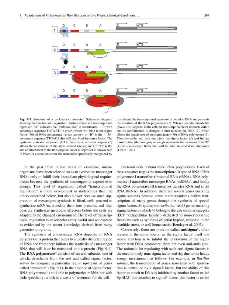
RNA that will later be translated into a protein (Fig. 9.1).
The RNA polymerase* consists of several subunits, one of
which, detachable from the rest and called sigma factor,
serves to recognize a particular region upstream of genes
called “promoter” (Fig. 9.1). In the absence of sigma factor,
RNA polymerase is still able to polymerize mRNA but with
little specificity, which is a waste of resources for the cell.
Bacterial cells contain three RNA polymerases. Each of
these enzymes targets the transcription of a type of RNA: RNA
polymerase I transcribes ribosomal RNA (rRNA), RNA polymerase II transcribes messenger RNAs (mRNAs), and finally
the RNA polymerase III transcribes transfer RNA and small
RNA (tRNA). In addition, there are several genes encoding
sigma subunits because some microorganisms realize transcription of many genes through the synthesis of special
sigma factors. Streptomyces coelicolor has 65 genes encoding
sigma factors of which 45 belong to the extracellular category
(ECF “extracellular family”) dedicated to non-cytoplasmic
functions such as synthesis of aerial hyphae, response to the
disulfide stress, or wall homeostasis (Bentley et al. 2002).
Conversely, there are proteins called antisigma*, often
present in the same operon as the sigma factor itself and
whose function is to inhibit the interaction of the sigma
factor with DNA promoters; there are even anti-antisigma.
The rationale for regulating with such anti-sigma factors is
the need to finely tune sigma factor activity due to the heavy
energy investment that follows. For example, in Bacillus
subtilis, the transcription of genes associated with sporulation is controlled by a sigmaF factor, but the ability of this
factor to attach to DNA is inhibited by another factor called
SpoIIAC that attaches to sigmaF factor; this factor is called
A
B
C
D
ATG
E
A
B
C
D
ATG
E
A
B
C
D
ATG
E
A
B
C
D
ATG
E
1
4
3
2
Fig. 9.1 Structure of a prokaryotic promoter. Schematic diagram
showing the function of a regulator, illustrated here is a transcriptional
repressor. “A” indicates the “Pribnow box” at coordinates À10, with
consensus sequence TATAAT (in green) which will bind to the sigma
factor (70) of RNA polymerase (green arrow); in “B” is the “À35”
consensus sequence TTGACA that will also bind the sigma factor. The
upstream activator sequence (UAS, “upstream activator sequence”)
allows the attachment of the alpha subunit (in red) in “C.” “D” is the
site of attachment to the transcription factor (a repressor is shown here
in blue). In a situation where the metabolite specifically recognized by
it is absent, the transcriptional repressor is bound to DNA and prevents
the insertion of the RNA polymerase (1). When a specific metabolite
(black oval) appears in the cell, the transcription factor interacts with it
and its conformation is changed; it then releases the DNA (2), which
allows the attachment of the sigma factor (70) of RNA polymerase (3).
Then the alpha and beta units join the sigma factor (3) and initiate
transcription (the dark gray rectangle represents the message) from “E”
(4) of a messenger RNA that will be later translated on ribosomes
(Lewin 1981)
9 Adaptations of Prokaryotes to Their Biotopes and to Physicochemical Conditions. . .
297
RNAs only to fulfill their immediate physiological requirements because the synthesis of messengers is expensive in
energy. This level of regulation, called “transcriptional
regulation,” is more economical in metabolites than the
others described below, but also slower because once suppression of messengers synthesis is lifted, cells proceed to
synthesize mRNAs, translate them into proteins, and then
possibly synthesize metabolic effectors before the cells are
adapted to the changed environment. The level of transcriptional regulation is nevertheless very useful and widespread
as evidenced by the recent knowledge derived from many
genomics programs.
The synthesis of a messenger RNA depends on RNA
polymerase, a protein that binds to a locally distorted region
of DNA and from there initiates the synthesis of a messenger
RNA that will later be translated into a protein (Fig. 9.1).
The RNA polymerase* consists of several subunits, one of
which, detachable from the rest and called sigma factor,
serves to recognize a particular region upstream of genes
called “promoter” (Fig. 9.1). In the absence of sigma factor,
RNA polymerase is still able to polymerize mRNA but with
little specificity, which is a waste of resources for the cell.
Bacterial cells contain three RNA polymerases. Each of
these enzymes targets the transcription of a type of RNA: RNA
polymerase I transcribes ribosomal RNA (rRNA), RNA polymerase II transcribes messenger RNAs (mRNAs), and finally
the RNA polymerase III transcribes transfer RNA and small
RNA (tRNA). In addition, there are several genes encoding
sigma subunits because some microorganisms realize transcription of many genes through the synthesis of special
sigma factors. Streptomyces coelicolor has 65 genes encoding
sigma factors of which 45 belong to the extracellular category
(ECF “extracellular family”) dedicated to non-cytoplasmic
functions such as synthesis of aerial hyphae, response to the
disulfide stress, or wall homeostasis (Bentley et al. 2002).
Conversely, there are proteins called antisigma*, often
present in the same operon as the sigma factor itself and
whose function is to inhibit the interaction of the sigma
factor with DNA promoters; there are even anti-antisigma.
The rationale for regulating with such anti-sigma factors is
the need to finely tune sigma factor activity due to the heavy
energy investment that follows. For example, in Bacillus
subtilis, the transcription of genes associated with sporulation is controlled by a sigmaF factor, but the ability of this
factor to attach to DNA is inhibited by another factor called
SpoIIAC that attaches to sigmaF factor; this factor is called
A
B
C
D
ATG
E
A
B
C
D
ATG
E
A
B
C
D
ATG
E
A
B
C
D
ATG
E
1
4
3
2
Fig. 9.1 Structure of a prokaryotic promoter. Schematic diagram
showing the function of a regulator, illustrated here is a transcriptional
repressor. “A” indicates the “Pribnow box” at coordinates À10, with
consensus sequence TATAAT (in green) which will bind to the sigma
factor (70) of RNA polymerase (green arrow); in “B” is the “À35”
consensus sequence TTGACA that will also bind the sigma factor. The
upstream activator sequence (UAS, “upstream activator sequence”)
allows the attachment of the alpha subunit (in red) in “C.” “D” is the
site of attachment to the transcription factor (a repressor is shown here
in blue). In a situation where the metabolite specifically recognized by
it is absent, the transcriptional repressor is bound to DNA and prevents
the insertion of the RNA polymerase (1). When a specific metabolite
(black oval) appears in the cell, the transcription factor interacts with it
and its conformation is changed; it then releases the DNA (2), which
allows the attachment of the sigma factor (70) of RNA polymerase (3).
Then the alpha and beta units join the sigma factor (3) and initiate
transcription (the dark gray rectangle represents the message) from “E”
(4) of a messenger RNA that will be later translated on ribosomes
(Lewin 1981)
9 Adaptations of Prokaryotes to Their Biotopes and to Physicochemical Conditions. . .
297