

1
Cyst
Cyst
Eukaryotic host
cell
Conidia
Nucleus
Polar
tube
Polar tube
Parasitic
Microsporidia
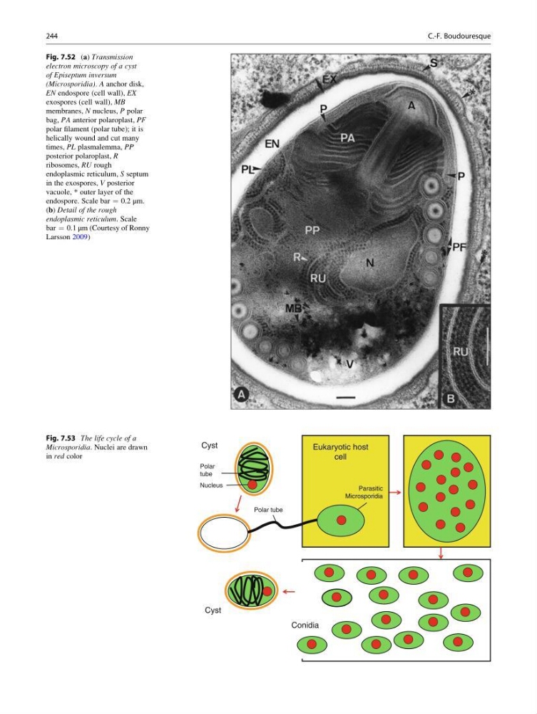
Fig. 7.53 The life cycle of a
Microsporidia. Nuclei are drawn
in red color
Fig. 7.52 (a) Transmission
electron microscopy of a cyst
of Episeptum inversum
(Microsporidia). A anchor disk,
EN endospore (cell wall), EX
exospores (cell wall), MB
membranes, N nucleus, P polar
bag, PA anterior polaroplast, PF
polar filament (polar tube); it is
helically wound and cut many
times, PL plasmalemma, PP
posterior polaroplast, R
ribosomes, RU rough
endoplasmic reticulum, S septum
in the exospores, V posterior
vacuole, * outer layer of the
endospore. Scale bar ¼ 0.2 μm.
(b) Detail of the rough
endoplasmic reticulum. Scale
bar ¼ 0.1 μm (Courtesy of Ronny
Larsson 2009)
244
C.-F. Boudouresque
Cyst
Cyst
Eukaryotic host
cell
Conidia
Nucleus
Polar
tube
Polar tube
Parasitic
Microsporidia
Fig. 7.53 The life cycle of a
Microsporidia. Nuclei are drawn
in red color
Fig. 7.52 (a) Transmission
electron microscopy of a cyst
of Episeptum inversum
(Microsporidia). A anchor disk,
EN endospore (cell wall), EX
exospores (cell wall), MB
membranes, N nucleus, P polar
bag, PA anterior polaroplast, PF
polar filament (polar tube); it is
helically wound and cut many
times, PL plasmalemma, PP
posterior polaroplast, R
ribosomes, RU rough
endoplasmic reticulum, S septum
in the exospores, V posterior
vacuole, * outer layer of the
endospore. Scale bar ¼ 0.2 μm.
(b) Detail of the rough
endoplasmic reticulum. Scale
bar ¼ 0.1 μm (Courtesy of Ronny
Larsson 2009)
244
C.-F. Boudouresque