

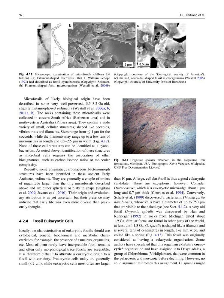
Microfossils of likely biological origin have been
described in some very well-preserved, 3.5–3.2-Ga-old,
slightly metamorphosed sediments (Westall et al. 2006a, b,
2011a, b). The rocks containing these microfossils were
collected in eastern South Africa (Barberton area) and in
northwestern Australia (Pilbara area). They contain a wide
variety of small, cellular structures, shaped like coccoids,
vibrios, rods and filaments. Sizes range from 1 μm for the
coccoids, while the filaments may range up to a few tens of
micrometres in length and 0.5–2.5 μm in width (Fig. 4.12).
None of these cell structures can be identified as a cyanobacterium. As noted above, identification of these structures
as microbial cells requires the association of other
biosignatures, such as carbon isotope ratios or molecular
complexity.
Recently, some enigmatic, carbonaceous bacteriomorph
structures have been identified in these ancient Early
Archaean sediments. They are generally a couple of orders
of magnitude larger than the tiny microfossils described
above and are either spherical or platy in shape (Sugitani
et al. 2009; Javaux et al. 2010). Their origin and evolutionary attribution is as yet uncertain, but their presence may
indicate that early life was even more diverse than previously thought.
4.2.4 Fossil Eukaryotic Cells
Ideally, the characterisation of eukaryotic fossils should use
cytological, genetic, biochemical and metabolic characteristics, for example, the presence of a nucleus, organelles,
etc. Most of them rarely leave interpretable fossil remains
and often only morphological trace fossils are accessible.
It is therefore difficult to attribute a eukaryotic origin to a
fossil with certainty. Prokaryotic cells today are generally
small (<2 μm), while eukaryotic cells most often are larger
than 10 μm. A large, cellular fossil is thus a good eukaryotic
candidate. There are exceptions, however. Consider
Ostreococcus, which is a eukaryotic micro-alga about 1 μm
long and 0.7 μm thick (Courties et al. 1994). Conversely,
Schulz et al. (1999) discovered a bacterium, Thiomargarita
namibiensis, whose cells have a diameter of up to 750 μm
that are visible to the naked eye (see Sect. 5.1.2). A very old
fossil Grypania spiralis was discovered by Han and
Runnegar (1992) in rocks from Michigan dated about
1.9 Ga. Similar forms are found in other parts of the world,
at least until 1.3 Ga. G. spiralis is shaped like a filament and
is several tens of centimetres in length, 1–2 mm wide, and
coiled like a spring (Fig. 4.13). This fossil was originally
considered as having a eukaryotic organisation. Some
authors have speculated that this organism exhibits a coenocytic* organisation and have assigned it to Dasycladales, a
group of Chlorobionta (Viridiplantae), that were common in
the palaeozoic and mesozoic before declining. However, no
solid argument reinforces this assignment. G. spiralis might
Fig. 4.12 Microscopic examination of microfossils (Pilbara 3.4
billion). (a) Filament-shaped microfossil that J. William Schopf
(1993) had described as fossil cyanobacteria (Copyright: Science).
(b) Filament-shaped fossil microorganism (Westall et al. 2006b)
(Copyright: courtesy of the ‘Geological Society of America’).
(c) chained, coccoidal-shaped fossil microorganisms (Westall 2005)
(Copyright: courtesy of University Press of Bordeaux)
Fig. 4.13 Grypania spiralis observed in the Negaunee iron
formations, Michigan, USA (Photographs: Xavie Vazquez, Wikipedia,
GNU Free Documentation Licence)
92
J.-C. Bertrand et al.
described in some very well-preserved, 3.5–3.2-Ga-old,
slightly metamorphosed sediments (Westall et al. 2006a, b,
2011a, b). The rocks containing these microfossils were
collected in eastern South Africa (Barberton area) and in
northwestern Australia (Pilbara area). They contain a wide
variety of small, cellular structures, shaped like coccoids,
vibrios, rods and filaments. Sizes range from 1 μm for the
coccoids, while the filaments may range up to a few tens of
micrometres in length and 0.5–2.5 μm in width (Fig. 4.12).
None of these cell structures can be identified as a cyanobacterium. As noted above, identification of these structures
as microbial cells requires the association of other
biosignatures, such as carbon isotope ratios or molecular
complexity.
Recently, some enigmatic, carbonaceous bacteriomorph
structures have been identified in these ancient Early
Archaean sediments. They are generally a couple of orders
of magnitude larger than the tiny microfossils described
above and are either spherical or platy in shape (Sugitani
et al. 2009; Javaux et al. 2010). Their origin and evolutionary attribution is as yet uncertain, but their presence may
indicate that early life was even more diverse than previously thought.
4.2.4 Fossil Eukaryotic Cells
Ideally, the characterisation of eukaryotic fossils should use
cytological, genetic, biochemical and metabolic characteristics, for example, the presence of a nucleus, organelles,
etc. Most of them rarely leave interpretable fossil remains
and often only morphological trace fossils are accessible.
It is therefore difficult to attribute a eukaryotic origin to a
fossil with certainty. Prokaryotic cells today are generally
small (<2 μm), while eukaryotic cells most often are larger
than 10 μm. A large, cellular fossil is thus a good eukaryotic
candidate. There are exceptions, however. Consider
Ostreococcus, which is a eukaryotic micro-alga about 1 μm
long and 0.7 μm thick (Courties et al. 1994). Conversely,
Schulz et al. (1999) discovered a bacterium, Thiomargarita
namibiensis, whose cells have a diameter of up to 750 μm
that are visible to the naked eye (see Sect. 5.1.2). A very old
fossil Grypania spiralis was discovered by Han and
Runnegar (1992) in rocks from Michigan dated about
1.9 Ga. Similar forms are found in other parts of the world,
at least until 1.3 Ga. G. spiralis is shaped like a filament and
is several tens of centimetres in length, 1–2 mm wide, and
coiled like a spring (Fig. 4.13). This fossil was originally
considered as having a eukaryotic organisation. Some
authors have speculated that this organism exhibits a coenocytic* organisation and have assigned it to Dasycladales, a
group of Chlorobionta (Viridiplantae), that were common in
the palaeozoic and mesozoic before declining. However, no
solid argument reinforces this assignment. G. spiralis might
Fig. 4.12 Microscopic examination of microfossils (Pilbara 3.4
billion). (a) Filament-shaped microfossil that J. William Schopf
(1993) had described as fossil cyanobacteria (Copyright: Science).
(b) Filament-shaped fossil microorganism (Westall et al. 2006b)
(Copyright: courtesy of the ‘Geological Society of America’).
(c) chained, coccoidal-shaped fossil microorganisms (Westall 2005)
(Copyright: courtesy of University Press of Bordeaux)
Fig. 4.13 Grypania spiralis observed in the Negaunee iron
formations, Michigan, USA (Photographs: Xavie Vazquez, Wikipedia,
GNU Free Documentation Licence)
92
J.-C. Bertrand et al.