

326
Edwardsiella ictaluri
Characteristics of the Disease
Enteric septicaemia of catfi sh was initially recognised in 1976 among populations
of pond-reared fi ngerlings and yearling fi sh in Alabama and Georgia, USA (Hawke
1979 ). Later, reports described its presence in Mississippi, Arkansas, Idaho,
Colorado, Indiana and Maryland. Although Edw. ictaluri is primarily a disease of
catfi sh, with time other fi sh groups have been reported to become infected, for
example, brown bullhead ( Amieurus nebulosus ) in the USA (Iwanowicz et al. 2006 )
and wild ayu ( Plecoglossus altivelis ) in Japan, particularly in September and
October when the temperature declined and the fi sh were sexually mature (Hassan
et al. 2012 ). The disease has spread from its original focal point in the USA to
include cultured striped catfi sh ( Pangasius hypophthalmus ) in Sumatra, Indonesia
(Yuasa et al. 2003 ) and catfi sh in Vietnam (Fig. 6.1 ; Crumlish et al. 2002 ), yellow
catfi sh ( Pelteobagrus fulvidraco ) (Ye et al. 2009 ) and southern catfi sh ( Silurus soldatovi meridionalis ) in China (Geng et al. 2013 ) and ayu in Japan (Sakai et al.
2008 ). It is apparent that there is a great variability in the clinical signs associated
with the disease. Just prior to death, the fi sh may hang listlessly in an almost vertical
position at the water surface, spin rapidly in circles, or exhibit spiral swimming.
External signs, often absent in fi sh over 15 cm in length, include the presence of
petechial (pin- prick) haemorrhages on the skin in the vicinity of the throat and
mouth, pale gills, exophthalmia and open lesions on the head, particularly on the
frontal bone of the skull between the eyes and on the lateral body surface. Internally,
there may be swelling of the kidney and spleen, haemorrhaging and necrotic areas
in the liver, blood-fi lled ascitic fl uid in the peritoneum, and petechial haemorrhages
throughout the internal muscle walls (Hawke 1979 ). In Vietnam, the disease is
referred to as “bacillary necrosis of Pangasius ” with which there are irregular white
lesions on the kidney, liver and spleen, and the involvement of parasites and bacteria
including Bacillus and Edw. ictaluri (Crumlish et al. 2002 ). With wild ayu, mortalities occurred from August to early October in 2007, with haemorrhagic ascites as a
common disease sign. Some fi sh demonstrated exophthalmia, and reddening along
the body surface, anus or the base of fi ns (Sakai et al. 2008 ).
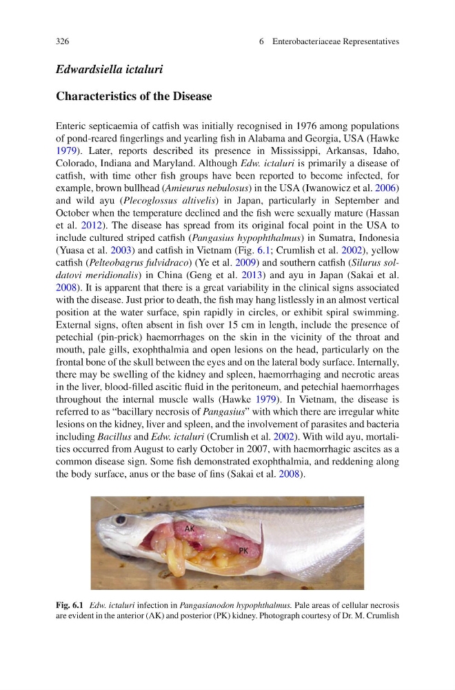
Fig. 6.1 Edw. ictaluri infection in Pangasianodon hypophthalmus. Pale areas of cellular necrosis
are evident in the anterior (AK) and posterior (PK) kidney. Photograph courtesy of Dr. M. Crumlish
6 Enterobacteriaceae Representatives
Edwardsiella ictaluri
Characteristics of the Disease
Enteric septicaemia of catfi sh was initially recognised in 1976 among populations
of pond-reared fi ngerlings and yearling fi sh in Alabama and Georgia, USA (Hawke
1979 ). Later, reports described its presence in Mississippi, Arkansas, Idaho,
Colorado, Indiana and Maryland. Although Edw. ictaluri is primarily a disease of
catfi sh, with time other fi sh groups have been reported to become infected, for
example, brown bullhead ( Amieurus nebulosus ) in the USA (Iwanowicz et al. 2006 )
and wild ayu ( Plecoglossus altivelis ) in Japan, particularly in September and
October when the temperature declined and the fi sh were sexually mature (Hassan
et al. 2012 ). The disease has spread from its original focal point in the USA to
include cultured striped catfi sh ( Pangasius hypophthalmus ) in Sumatra, Indonesia
(Yuasa et al. 2003 ) and catfi sh in Vietnam (Fig. 6.1 ; Crumlish et al. 2002 ), yellow
catfi sh ( Pelteobagrus fulvidraco ) (Ye et al. 2009 ) and southern catfi sh ( Silurus soldatovi meridionalis ) in China (Geng et al. 2013 ) and ayu in Japan (Sakai et al.
2008 ). It is apparent that there is a great variability in the clinical signs associated
with the disease. Just prior to death, the fi sh may hang listlessly in an almost vertical
position at the water surface, spin rapidly in circles, or exhibit spiral swimming.
External signs, often absent in fi sh over 15 cm in length, include the presence of
petechial (pin- prick) haemorrhages on the skin in the vicinity of the throat and
mouth, pale gills, exophthalmia and open lesions on the head, particularly on the
frontal bone of the skull between the eyes and on the lateral body surface. Internally,
there may be swelling of the kidney and spleen, haemorrhaging and necrotic areas
in the liver, blood-fi lled ascitic fl uid in the peritoneum, and petechial haemorrhages
throughout the internal muscle walls (Hawke 1979 ). In Vietnam, the disease is
referred to as “bacillary necrosis of Pangasius ” with which there are irregular white
lesions on the kidney, liver and spleen, and the involvement of parasites and bacteria
including Bacillus and Edw. ictaluri (Crumlish et al. 2002 ). With wild ayu, mortalities occurred from August to early October in 2007, with haemorrhagic ascites as a
common disease sign. Some fi sh demonstrated exophthalmia, and reddening along
the body surface, anus or the base of fi ns (Sakai et al. 2008 ).
Fig. 6.1 Edw. ictaluri infection in Pangasianodon hypophthalmus. Pale areas of cellular necrosis
are evident in the anterior (AK) and posterior (PK) kidney. Photograph courtesy of Dr. M. Crumlish
6 Enterobacteriaceae Representatives