

86
and histiocytes, degenerating leucocytes with macrophages, and fi brin (Wolke
1975 ). In Atlantic salmon, petechial haemorrhages have been described on the muscle lining the peritoneum (Smith 1964 ). However, Ren. salmoninarum has been
found in salmonids, e.g. char and grayling in Alaska, without any evidence of clinical disease (Meyers et al. 1993 ). Renibacterial cells have been found in intraabdominal adhesions in Atlantic salmon and coho salmon that may have previously
or were considered to have received adjuvant (= adjuvanted vaccine) intraperitoneally (Bruno and Brown 1999 ). Evidence suggests an impact of the disease on appetite with infected fi sh consuming less than their less diseased counterparts (Pirhonen
et al. 2000 ). A reduction in growth of infect Chinook salmon has been observed
(Sandell et al. 2015 ). One point that needs to be emphasised is that BKD does not
always manifest itself in a clinical form, sub-clinical disease poses a real risk of
transfer of the pathogen with fi sh movements, especially if the animals are perceived to be healthy (Murray et al. 2011 ).
Isolation
Cultivation of the causal agent of BKD in vitro from chinook salmon was not
achieved until Earp ( 1950 ) used a nutrient-rich medium, containing fi sh extract,
glucose, yeast extract and bovine serum/meat infusion, with incubation at 15 or
20 °C. An improvement resulted from use of minced chick embryos in 1 % (w/v)
agar or on Dorset egg medium (Earp et al. 1953 ). Nevertheless, growth was generally poor, even after prolonged incubation periods of ≥14 days. To prove that the
growth was of the pathogen, Earp successfully inoculated the bacterial culture into
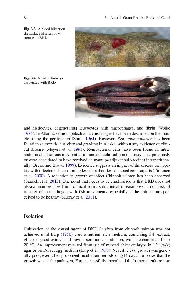
Fig. 3.3 A blood blister on
the surface of a rainbow
trout with BKD
Fig. 3.4 Swollen kidneys
associated with BKD
3 Aerobic Gram-Positive Rods and Cocci
and histiocytes, degenerating leucocytes with macrophages, and fi brin (Wolke
1975 ). In Atlantic salmon, petechial haemorrhages have been described on the muscle lining the peritoneum (Smith 1964 ). However, Ren. salmoninarum has been
found in salmonids, e.g. char and grayling in Alaska, without any evidence of clinical disease (Meyers et al. 1993 ). Renibacterial cells have been found in intraabdominal adhesions in Atlantic salmon and coho salmon that may have previously
or were considered to have received adjuvant (= adjuvanted vaccine) intraperitoneally (Bruno and Brown 1999 ). Evidence suggests an impact of the disease on appetite with infected fi sh consuming less than their less diseased counterparts (Pirhonen
et al. 2000 ). A reduction in growth of infect Chinook salmon has been observed
(Sandell et al. 2015 ). One point that needs to be emphasised is that BKD does not
always manifest itself in a clinical form, sub-clinical disease poses a real risk of
transfer of the pathogen with fi sh movements, especially if the animals are perceived to be healthy (Murray et al. 2011 ).
Isolation
Cultivation of the causal agent of BKD in vitro from chinook salmon was not
achieved until Earp ( 1950 ) used a nutrient-rich medium, containing fi sh extract,
glucose, yeast extract and bovine serum/meat infusion, with incubation at 15 or
20 °C. An improvement resulted from use of minced chick embryos in 1 % (w/v)
agar or on Dorset egg medium (Earp et al. 1953 ). Nevertheless, growth was generally poor, even after prolonged incubation periods of ≥14 days. To prove that the
growth was of the pathogen, Earp successfully inoculated the bacterial culture into
Fig. 3.3 A blood blister on
the surface of a rainbow
trout with BKD
Fig. 3.4 Swollen kidneys
associated with BKD
3 Aerobic Gram-Positive Rods and Cocci