

424
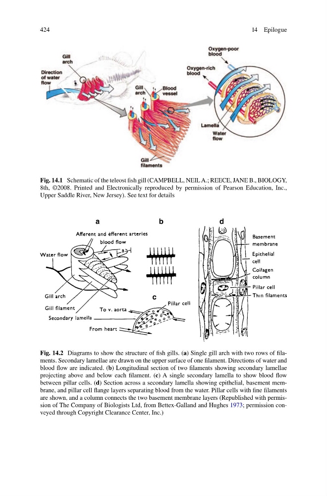
Fig. 14.1 Schematic of the teleost fi sh gill (CAMPBELL, NEIL A.; REECE, JANE B., BIOLOGY,
8th, ©2008. Printed and Electronically reproduced by permission of Pearson Education, Inc.,
Upper Saddle River, New Jersey). See text for details
Fig. 14.2 Diagrams to show the structure of fi sh gills. ( a ) Single gill arch with two rows of fi laments. Secondary lamellae are drawn on the upper surface of one fi lament. Directions of water and
blood fl ow are indicated. ( b ) Longitudinal section of two fi laments showing secondary lamellae
projecting above and below each fi lament. ( c ) A single secondary lamella to show blood fl ow
between pillar cells. ( d ) Section across a secondary lamella showing epithelial, basement membrane, and pillar cell fl ange layers separating blood from the water. Pillar cells with fi ne fi laments
are shown, and a column connects the two basement membrane layers (Republished with permission of The Company of Biologists Ltd, from Bettex-Galland and Hughes 1973 ; permission conveyed through Copyright Clearance Center, Inc.)
14 Epilogue
Fig. 14.1 Schematic of the teleost fi sh gill (CAMPBELL, NEIL A.; REECE, JANE B., BIOLOGY,
8th, ©2008. Printed and Electronically reproduced by permission of Pearson Education, Inc.,
Upper Saddle River, New Jersey). See text for details
Fig. 14.2 Diagrams to show the structure of fi sh gills. ( a ) Single gill arch with two rows of fi laments. Secondary lamellae are drawn on the upper surface of one fi lament. Directions of water and
blood fl ow are indicated. ( b ) Longitudinal section of two fi laments showing secondary lamellae
projecting above and below each fi lament. ( c ) A single secondary lamella to show blood fl ow
between pillar cells. ( d ) Section across a secondary lamella showing epithelial, basement membrane, and pillar cell fl ange layers separating blood from the water. Pillar cells with fi ne fi laments
are shown, and a column connects the two basement membrane layers (Republished with permission of The Company of Biologists Ltd, from Bettex-Galland and Hughes 1973 ; permission conveyed through Copyright Clearance Center, Inc.)
14 Epilogue