

227
The thin layer of monocrystalline enameloid is located on the surface of each scale.
Some scales (pore-scales) of the Thelodontian Phlebolepis elegans are penetrated
by one or more channels which extend on the inner side of the scales in the shape of
tubes. The position and number of these channels is quite arbitrary. The channels of
these pore-scales are interpreted as branch channels of the lateral organ (Gross
1967 ).
Elasmoid Scales The adjective “ elasmoid ” has been coined by Bertin ( 1944 ). These
scales represent commonest type of scale, they are thin and fl exible as well as possess
typical lamellar structure and imbricated fi tting in the tegument. This is the difference to the rhomboid scale arrangement of “lower” bony fi shes (see Goodrich 1907 ).
This defi nition allows the classifi cation of structures as elasmoid scales of the living
Coelacanthidae ( Latimeria ), Dipnoi ( Lepidosiren , Neoceratodus, Protopterus ), and
Amiidae ( Amid ) (Meunier 1983 ). Structural features of elasmoid scales has been
described in several species belonging to the sarcopterygian and the actinopterygian
lineages (see Meunier 1983 , 1984 ; Sire 1987 ; Huysseune and Sire 1998 ). Most of the
26,000 Teleostei species possess elasmoid scales, which forms in the fi sh dermis
without the presence of a cartilaginous initium (see for review Francillon-Vieillot
et al. 1990 ; Zylberberg et al. 1992 ). Sire and Akimenko ( 2004 ) reported that the elasmoid scales were mostly found to be invariably composed of three tissues:
(1) “the basal plate, a thick layer of incompletely mineralised tissue composed of
elasmodin (previously called isopedin), itself consisting of several layers of
type I and V collagen fi brils organised into a plywood-like structure (Meunier
1983 ; Schultze 1996 );
(2) the external layer, a thin layer of well-mineralised tissue composed of a network
of interwoven collagen fi brils;
(3) the limiting layer, a hyper-mineralised tissue devoid of collagen fi brils and
deposited at the scale surface in the region close to the epidermis. The structure
and organisation of this upper layer is the most variable amongst the various
species,” (Sire and Akimenko 2004 ).
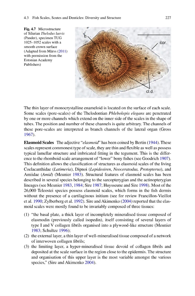
Fig. 4.7 Microstructure
of Silurian Thelodus laevis
(Pander), specimen TUG
1025–1052 scales with a
smooth crown surface
(Adapted from Märss ( 2011 )
with permission from the
Estonian Academy
Publishers)
4.3 Fish Scales, Scutes and Denticles: Diversity and Structure
The thin layer of monocrystalline enameloid is located on the surface of each scale.
Some scales (pore-scales) of the Thelodontian Phlebolepis elegans are penetrated
by one or more channels which extend on the inner side of the scales in the shape of
tubes. The position and number of these channels is quite arbitrary. The channels of
these pore-scales are interpreted as branch channels of the lateral organ (Gross
1967 ).
Elasmoid Scales The adjective “ elasmoid ” has been coined by Bertin ( 1944 ). These
scales represent commonest type of scale, they are thin and fl exible as well as possess
typical lamellar structure and imbricated fi tting in the tegument. This is the difference to the rhomboid scale arrangement of “lower” bony fi shes (see Goodrich 1907 ).
This defi nition allows the classifi cation of structures as elasmoid scales of the living
Coelacanthidae ( Latimeria ), Dipnoi ( Lepidosiren , Neoceratodus, Protopterus ), and
Amiidae ( Amid ) (Meunier 1983 ). Structural features of elasmoid scales has been
described in several species belonging to the sarcopterygian and the actinopterygian
lineages (see Meunier 1983 , 1984 ; Sire 1987 ; Huysseune and Sire 1998 ). Most of the
26,000 Teleostei species possess elasmoid scales, which forms in the fi sh dermis
without the presence of a cartilaginous initium (see for review Francillon-Vieillot
et al. 1990 ; Zylberberg et al. 1992 ). Sire and Akimenko ( 2004 ) reported that the elasmoid scales were mostly found to be invariably composed of three tissues:
(1) “the basal plate, a thick layer of incompletely mineralised tissue composed of
elasmodin (previously called isopedin), itself consisting of several layers of
type I and V collagen fi brils organised into a plywood-like structure (Meunier
1983 ; Schultze 1996 );
(2) the external layer, a thin layer of well-mineralised tissue composed of a network
of interwoven collagen fi brils;
(3) the limiting layer, a hyper-mineralised tissue devoid of collagen fi brils and
deposited at the scale surface in the region close to the epidermis. The structure
and organisation of this upper layer is the most variable amongst the various
species,” (Sire and Akimenko 2004 ).
Fig. 4.7 Microstructure
of Silurian Thelodus laevis
(Pander), specimen TUG
1025–1052 scales with a
smooth crown surface
(Adapted from Märss ( 2011 )
with permission from the
Estonian Academy
Publishers)
4.3 Fish Scales, Scutes and Denticles: Diversity and Structure