

180
weak electrical discharges to the surrounding environment, are the place for silica
localization. These organs possess specialized cells known as electrocytes (Prado
Figueroa 2005 ; Prado Figueroa et al. 2008 ; Prado Figueroa 2011 ). They were
described as follow: “Electrocytes are cup-shaped cells, highly polarized, with
an anterior, concave, innervated face and a posterior, convex, noninnervated face
that shows a very large system of caveolae,” (Prado Figueroa et al. 2008 ). Based
on the evidence of aluminium (Barrera et al. 2001 ) and silicon accumulation in
electrocytes from P. extenta by SEM and energy dispersive X-ray microanalysis
(EDS), Prado Figueroa and co-workers recently reported the presence of silica in
the form of chalcedony. This crystalline mineral was detected in the cytoplasm
and synaptic regions of electrocytes using a mineralogical microscope. The
localization of silica in the subcellular fractions of electric organs of P. extenta
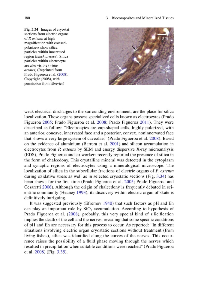
during oxidative stress as well as in selected cryostatic sections (Fig. 3.34 ) has
been shown for the fi rst time (Prado Figueroa et al. 2005 ; Prado Figueroa and
Cesaretti 2006 ). Although the origin of chalcedony is frequently debated in scientifi c community (Heaney 1993 ), its discovery within electric organ of skate is
defi nitively intriguing.
It was suggested previously (Efremov 1940 ) that such factors as pH and Eh
can play an important role by SiO 2 accumulation. According to hypothesis of
Prado Figueroa et al. ( 2008 ), probably, this very special kind of silicifi cation
implies the death of the cell and the nerves, revealing that some specifi c conditions
of pH and Eh are necessary for this process to occur. As reported: “In different
situations involving electric organ cryostatic sections without treatment (from
living fi shes), silica was identifi ed along the curves of the nerves. This occurrence raises the possibility of a fl uid phase moving through the nerves which
resulted in precipitation when suitable conditions were reached” (Prado Figueroa
et al. 2008 ) (Fig. 3.35 ).
Fig. 3.34 Images of cryostat
sections from electric organs
of P. extenta at high
magnifi cation with crossedpolarizers show silica
particles within innervated
region ( black arrows ). Silica
particles within electrocyte
are also visible ( white
arrows ) (Reprinted from
Prado Figueroa et al. ( 2008 ),
Copyright (2008), with
permission from Elsevier)
3 Biocomposites and Mineralized Tissues
weak electrical discharges to the surrounding environment, are the place for silica
localization. These organs possess specialized cells known as electrocytes (Prado
Figueroa 2005 ; Prado Figueroa et al. 2008 ; Prado Figueroa 2011 ). They were
described as follow: “Electrocytes are cup-shaped cells, highly polarized, with
an anterior, concave, innervated face and a posterior, convex, noninnervated face
that shows a very large system of caveolae,” (Prado Figueroa et al. 2008 ). Based
on the evidence of aluminium (Barrera et al. 2001 ) and silicon accumulation in
electrocytes from P. extenta by SEM and energy dispersive X-ray microanalysis
(EDS), Prado Figueroa and co-workers recently reported the presence of silica in
the form of chalcedony. This crystalline mineral was detected in the cytoplasm
and synaptic regions of electrocytes using a mineralogical microscope. The
localization of silica in the subcellular fractions of electric organs of P. extenta
during oxidative stress as well as in selected cryostatic sections (Fig. 3.34 ) has
been shown for the fi rst time (Prado Figueroa et al. 2005 ; Prado Figueroa and
Cesaretti 2006 ). Although the origin of chalcedony is frequently debated in scientifi c community (Heaney 1993 ), its discovery within electric organ of skate is
defi nitively intriguing.
It was suggested previously (Efremov 1940 ) that such factors as pH and Eh
can play an important role by SiO 2 accumulation. According to hypothesis of
Prado Figueroa et al. ( 2008 ), probably, this very special kind of silicifi cation
implies the death of the cell and the nerves, revealing that some specifi c conditions
of pH and Eh are necessary for this process to occur. As reported: “In different
situations involving electric organ cryostatic sections without treatment (from
living fi shes), silica was identifi ed along the curves of the nerves. This occurrence raises the possibility of a fl uid phase moving through the nerves which
resulted in precipitation when suitable conditions were reached” (Prado Figueroa
et al. 2008 ) (Fig. 3.35 ).
Fig. 3.34 Images of cryostat
sections from electric organs
of P. extenta at high
magnifi cation with crossedpolarizers show silica
particles within innervated
region ( black arrows ). Silica
particles within electrocyte
are also visible ( white
arrows ) (Reprinted from
Prado Figueroa et al. ( 2008 ),
Copyright (2008), with
permission from Elsevier)
3 Biocomposites and Mineralized Tissues