

107
defi nitions, odontodes are “ all structures that comprise a mineralized hard tissue
unit consisting of attachment bony basal pad, dentine or similar dentinous tissue.
Occasionally there is a superfi cial layer of enamel/enameloid formed from a single
papilla; odontodes include both teeth and denticles, ” (Fraser et al. 2010 ). Principal
question concerning the appearance and development of odontodes during early
vertebrate evolution is logically rooted in the classic problem of anatomical homology
(Ørvig 1977 ). There are numerous locations around the body of lower vertebrates, where
odontodes can be observed. For example, covering the dermal surface as in extant
rays and sharks, as lining the oropharyngeal cavity associated with gill arches, or
dentition in oral and pharyngeal locations (Sire and Huysseune 2003 ). The perceived
homology of all odontodes is suggested using palaeontological data, comparing
developmental and structural similarities (see for review Fraser et al. 2010 ).
Location, morphology and function of odontodes even within one organism can
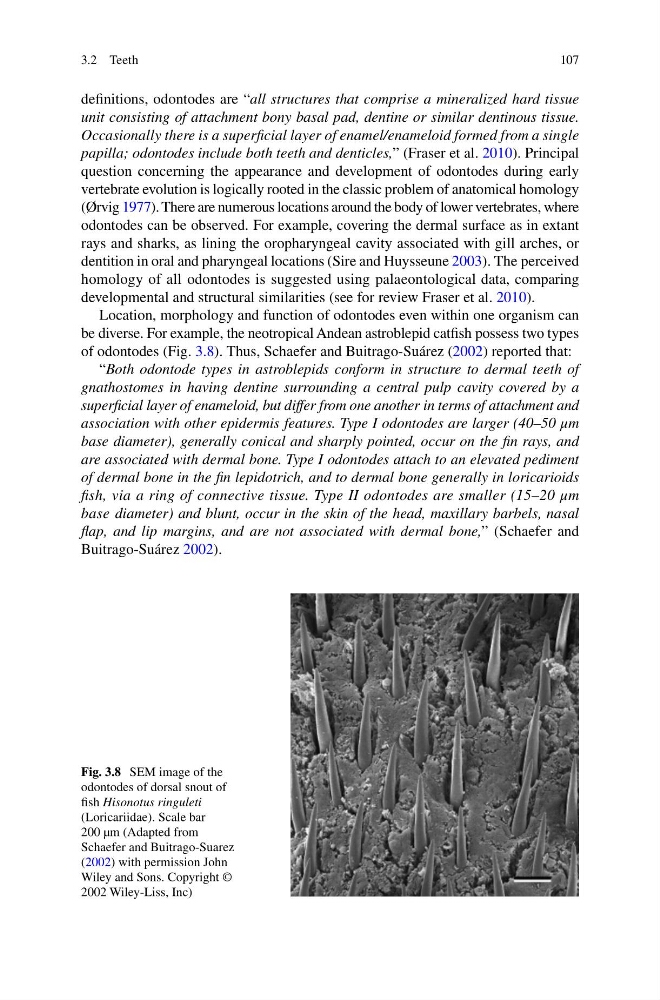
be diverse. For example, the neotropical Andean astroblepid catfi sh possess two types
of odontodes (Fig. 3.8 ). Thus, Schaefer and Buitrago-Suárez ( 2002 ) reported that:
“ Both odontode types in astroblepids conform in structure to dermal teeth of
gnathostomes in having dentine surrounding a central pulp cavity covered by a
superfi cial layer of enameloid, but differ from one another in terms of attachment and
association with other epidermis features. Type I odontodes are larger (40–50 μm
base diameter), generally conical and sharply pointed, occur on the fi n rays, and
are associated with dermal bone. Type I odontodes attach to an elevated pediment
of dermal bone in the fi n lepidotrich, and to dermal bone generally in loricarioids
fi sh, via a ring of connective tissue. Type II odontodes are smaller (15–20 μm
base diameter) and blunt, occur in the skin of the head, maxillary barbels, nasal
fl ap, and lip margins, and are not associated with dermal bone, ” (Schaefer and
Buitrago-Suárez 2002 ).
Fig. 3.8 SEM image of the
odontodes of dorsal snout of
fi sh Hisonotus ringuleti
(Loricariidae). Scale bar
200 µm (Adapted from
Schaefer and Buitrago-Suarez
( 2002 ) with permission John
Wiley and Sons. Copyright ©
2002 Wiley-Liss, Inc)
3.2 Teeth
defi nitions, odontodes are “ all structures that comprise a mineralized hard tissue
unit consisting of attachment bony basal pad, dentine or similar dentinous tissue.
Occasionally there is a superfi cial layer of enamel/enameloid formed from a single
papilla; odontodes include both teeth and denticles, ” (Fraser et al. 2010 ). Principal
question concerning the appearance and development of odontodes during early
vertebrate evolution is logically rooted in the classic problem of anatomical homology
(Ørvig 1977 ). There are numerous locations around the body of lower vertebrates, where
odontodes can be observed. For example, covering the dermal surface as in extant
rays and sharks, as lining the oropharyngeal cavity associated with gill arches, or
dentition in oral and pharyngeal locations (Sire and Huysseune 2003 ). The perceived
homology of all odontodes is suggested using palaeontological data, comparing
developmental and structural similarities (see for review Fraser et al. 2010 ).
Location, morphology and function of odontodes even within one organism can
be diverse. For example, the neotropical Andean astroblepid catfi sh possess two types
of odontodes (Fig. 3.8 ). Thus, Schaefer and Buitrago-Suárez ( 2002 ) reported that:
“ Both odontode types in astroblepids conform in structure to dermal teeth of
gnathostomes in having dentine surrounding a central pulp cavity covered by a
superfi cial layer of enameloid, but differ from one another in terms of attachment and
association with other epidermis features. Type I odontodes are larger (40–50 μm
base diameter), generally conical and sharply pointed, occur on the fi n rays, and
are associated with dermal bone. Type I odontodes attach to an elevated pediment
of dermal bone in the fi n lepidotrich, and to dermal bone generally in loricarioids
fi sh, via a ring of connective tissue. Type II odontodes are smaller (15–20 μm
base diameter) and blunt, occur in the skin of the head, maxillary barbels, nasal
fl ap, and lip margins, and are not associated with dermal bone, ” (Schaefer and
Buitrago-Suárez 2002 ).
Fig. 3.8 SEM image of the
odontodes of dorsal snout of
fi sh Hisonotus ringuleti
(Loricariidae). Scale bar
200 µm (Adapted from
Schaefer and Buitrago-Suarez
( 2002 ) with permission John
Wiley and Sons. Copyright ©
2002 Wiley-Liss, Inc)
3.2 Teeth