

recombinant ferritins identical except for the alanine/serine shift, have significant
effects on K cat (Tosha et al. 2008). Thus, while the sequence motifs for Fe 2 in both
mini-ferritin and bacterioferritin consist of a few common motifs found in the
majority of the ferritins, the variations could have functional significance.
2.3 Ferritin Iron Biomineral Formation
Iron biomineralization in ferritins is a multistage process with discretely defined
steps such as ferrous substrate binding, oxidation, product (mineral precursor)
release and, when the active sites are in the middle of the protein cage, mineral
nucleation, as recently observed by 13C–13C and magnetic susceptibility NMR
(Turano et al. 2010). The multiphase process is illustrated in progress curves where
data are collected at a general wavelength for Fe(III)O species (310–420 nm) as in
Fig. 2.3. By contrast, when data are collected at the lmax of the differic peroxo
complex (650 nm), a single phase of the reaction is observed (Fig. 2.3).
A goal for the future is deconvolution, both spectroscopic and kinetic, of the
multiple intermediates in ferritin iron biomineralization. The different steps in
ferritin biomineral synthesis are:
1. Fe (II) enters the protein cage and binds at the active sites
2. Oxygen or hydrogen peroxide binds with oxidoreduction and formation of
transition intermediates
3. Release of ferric oxo mineral precursor
4. Nucleation and mineralization
2.3.1 Step i. Fe(II) Entry and Binding
Binding of Fe(II) to the active site has been studied directly by MCD/CD in a
eukaryotic maxi-ferritin (Schwartz et al. 2008), and indirectly by soaking Fe(II)
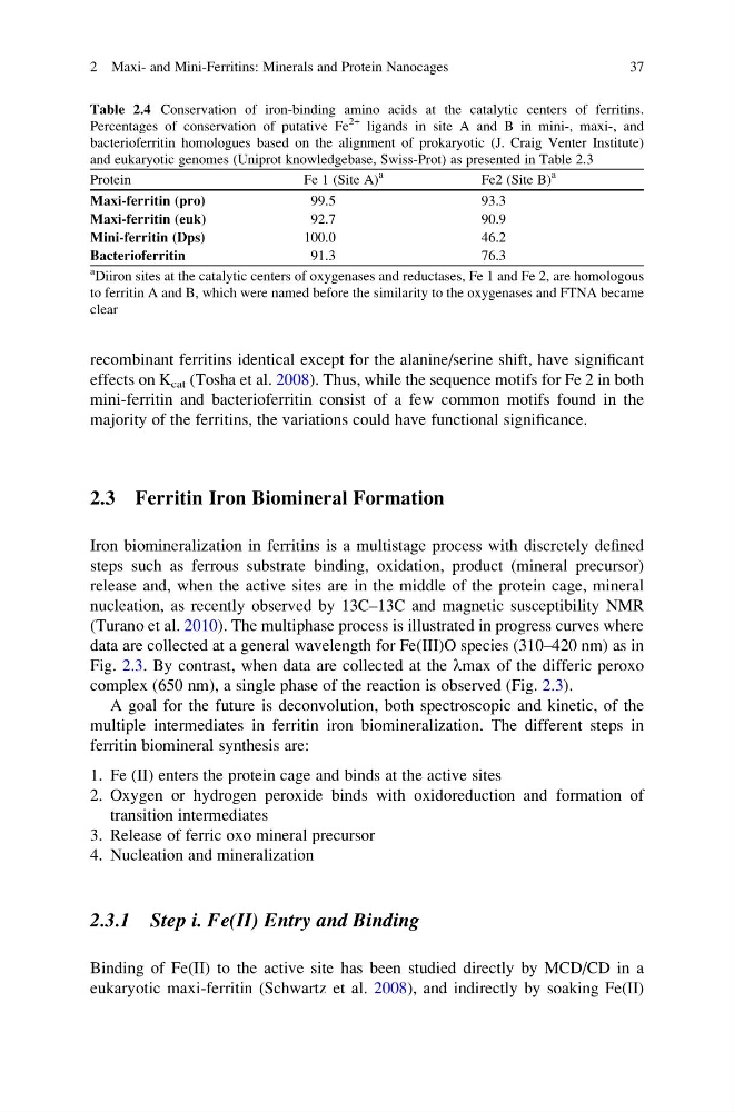
Table 2.4 Conservation of iron-binding amino acids at the catalytic centers of ferritins.
Percentages of conservation of putative Fe
2+ ligands in site A and B in mini-, maxi-, and
bacterioferritin homologues based on the alignment of prokaryotic (J. Craig Venter Institute)
and eukaryotic genomes (Uniprot knowledgebase, Swiss-Prot) as presented in Table 2.3
Protein
Fe 1 (Site A)
a
Fe2 (Site B)
a
Maxi-ferritin (pro)
99.5
93.3
Maxi-ferritin (euk)
92.7
90.9
Mini-ferritin (Dps)
100.0
46.2
Bacterioferritin
91.3
76.3
a
Diiron sites at the catalytic centers of oxygenases and reductases, Fe 1 and Fe 2, are homologous
to ferritin A and B, which were named before the similarity to the oxygenases and FTNA became
clear
2 Maxi- and Mini-Ferritins: Minerals and Protein Nanocages
37
effects on K cat (Tosha et al. 2008). Thus, while the sequence motifs for Fe 2 in both
mini-ferritin and bacterioferritin consist of a few common motifs found in the
majority of the ferritins, the variations could have functional significance.
2.3 Ferritin Iron Biomineral Formation
Iron biomineralization in ferritins is a multistage process with discretely defined
steps such as ferrous substrate binding, oxidation, product (mineral precursor)
release and, when the active sites are in the middle of the protein cage, mineral
nucleation, as recently observed by 13C–13C and magnetic susceptibility NMR
(Turano et al. 2010). The multiphase process is illustrated in progress curves where
data are collected at a general wavelength for Fe(III)O species (310–420 nm) as in
Fig. 2.3. By contrast, when data are collected at the lmax of the differic peroxo
complex (650 nm), a single phase of the reaction is observed (Fig. 2.3).
A goal for the future is deconvolution, both spectroscopic and kinetic, of the
multiple intermediates in ferritin iron biomineralization. The different steps in
ferritin biomineral synthesis are:
1. Fe (II) enters the protein cage and binds at the active sites
2. Oxygen or hydrogen peroxide binds with oxidoreduction and formation of
transition intermediates
3. Release of ferric oxo mineral precursor
4. Nucleation and mineralization
2.3.1 Step i. Fe(II) Entry and Binding
Binding of Fe(II) to the active site has been studied directly by MCD/CD in a
eukaryotic maxi-ferritin (Schwartz et al. 2008), and indirectly by soaking Fe(II)
Table 2.4 Conservation of iron-binding amino acids at the catalytic centers of ferritins.
Percentages of conservation of putative Fe
2+ ligands in site A and B in mini-, maxi-, and
bacterioferritin homologues based on the alignment of prokaryotic (J. Craig Venter Institute)
and eukaryotic genomes (Uniprot knowledgebase, Swiss-Prot) as presented in Table 2.3
Protein
Fe 1 (Site A)
a
Fe2 (Site B)
a
Maxi-ferritin (pro)
99.5
93.3
Maxi-ferritin (euk)
92.7
90.9
Mini-ferritin (Dps)
100.0
46.2
Bacterioferritin
91.3
76.3
a
Diiron sites at the catalytic centers of oxygenases and reductases, Fe 1 and Fe 2, are homologous
to ferritin A and B, which were named before the similarity to the oxygenases and FTNA became
clear
2 Maxi- and Mini-Ferritins: Minerals and Protein Nanocages
37