

2.2.1 Ferritin in Prokaryotes
Primary sequences for both maxi- and mini-ferritins are widespread in the genomes
of prokaryotes, and even within the genome of one single organism. E. coli is an
example of an organism that has gene copies for multiple ferritins. E. coli miniferritin (Dps), maxi-ferritin (FTNA), and bacterioferritin (BFR) were used as
queries in BLAST searches in order to study the distribution of recognizable
ferritins among prokaryotes (P-value <5 Â 10
À5 ), and to generate alignments
that provide insights into the conservation of active site residues. The term “recognizable” stresses the limitation of the approach to find homologous proteins based
on sequence homology on its own. In the case of ferritin in particular, the threedimensional protein structure is highly conserved and essential for its function.
However, there are no means yet to screen multiple genomes for the presence of
specific, quarternary, protein structures. The results of the sequence homology
studies cannot be the “final picture” of the ferritin family, because of sequencing
gaps. For example, sequenced archaeal genomes, to date, are 12-fold fewer than
sequence bacterial genomes (42 vs. 524). We only present a snapshot to illustrate
the broad distribution patterns of mini-ferritins (Dps), maxi-ferritins, and bacterioferritins (BFRs), and the conservation of the catalytic sites.
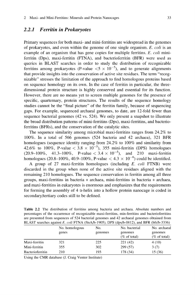
The sequence similarity among microbial maxi-ferritins ranges from 24.2% to
100%. In a total of 566 genomes (524 bacteria and 42 archaea), 321 BFR
homologues (sequence identity ranging from 24.2% to 100% and similarity from
42.6% to 100%, P-value < 3.8 Â 10
À5 ), 355 mini-ferritin (DPS) homologues
(20.9–100%, 41.3–100%, P-value < 3.4 Â 10
À5 ), and 210 maxi-ferritin
homologues (20.8–100%, 40.9–100%, P-value < 4.3 Â 10
À6 ) could be identified.
A group of 27 maxi-ferritin homologues (including E. coli FTNB) were
discarded in the group when none of the active site residues aligned with the
remaining 210 homologues. The sequence conservation in ferritin among all three
groups, maxi-ferritins in bacteria + archaea, mini-ferritins in bacteria + archaea,
and maxi-ferritins in eukaryotes is enormous and emphasizes that the requirements
for forming the assembly of 4 a-helix into a hollow protein nanocage is coded in
secondary/tertiary codes still to be defined.
Table 2.2 The distribution of ferritins among bacteria and archaea. Absolute numbers and
percentages of the occurrence of recognizable maxi-ferritins, min-ferritins and bacterioferritins
are presented from sequences of 524 bacterial genomes and 42 archaeal genomes obtained from
BLAST searches against E. coli FTNA (ftnA/b-1905), DPS (dps/b-0812), and BFR (bfr/b-3336)
No. homologous
genes
No.
genomes
No. bacterial
genomes
(% of total)
No. archaeal
genomes
(% of total)
Maxi-ferritin
321
225
221 (42)
4 (10)
Mini-ferritin
355
302
299 (57)
3 (7)
Bacterioferritin
210
193
178 (34)
15 (36)
Using the CMR database (J. Craig Venter Institute)
2 Maxi- and Mini-Ferritins: Minerals and Protein Nanocages
33
Primary sequences for both maxi- and mini-ferritins are widespread in the genomes
of prokaryotes, and even within the genome of one single organism. E. coli is an
example of an organism that has gene copies for multiple ferritins. E. coli miniferritin (Dps), maxi-ferritin (FTNA), and bacterioferritin (BFR) were used as
queries in BLAST searches in order to study the distribution of recognizable
ferritins among prokaryotes (P-value <5 Â 10
À5 ), and to generate alignments
that provide insights into the conservation of active site residues. The term “recognizable” stresses the limitation of the approach to find homologous proteins based
on sequence homology on its own. In the case of ferritin in particular, the threedimensional protein structure is highly conserved and essential for its function.
However, there are no means yet to screen multiple genomes for the presence of
specific, quarternary, protein structures. The results of the sequence homology
studies cannot be the “final picture” of the ferritin family, because of sequencing
gaps. For example, sequenced archaeal genomes, to date, are 12-fold fewer than
sequence bacterial genomes (42 vs. 524). We only present a snapshot to illustrate
the broad distribution patterns of mini-ferritins (Dps), maxi-ferritins, and bacterioferritins (BFRs), and the conservation of the catalytic sites.
The sequence similarity among microbial maxi-ferritins ranges from 24.2% to
100%. In a total of 566 genomes (524 bacteria and 42 archaea), 321 BFR
homologues (sequence identity ranging from 24.2% to 100% and similarity from
42.6% to 100%, P-value < 3.8 Â 10
À5 ), 355 mini-ferritin (DPS) homologues
(20.9–100%, 41.3–100%, P-value < 3.4 Â 10
À5 ), and 210 maxi-ferritin
homologues (20.8–100%, 40.9–100%, P-value < 4.3 Â 10
À6 ) could be identified.
A group of 27 maxi-ferritin homologues (including E. coli FTNB) were
discarded in the group when none of the active site residues aligned with the
remaining 210 homologues. The sequence conservation in ferritin among all three
groups, maxi-ferritins in bacteria + archaea, mini-ferritins in bacteria + archaea,
and maxi-ferritins in eukaryotes is enormous and emphasizes that the requirements
for forming the assembly of 4 a-helix into a hollow protein nanocage is coded in
secondary/tertiary codes still to be defined.
Table 2.2 The distribution of ferritins among bacteria and archaea. Absolute numbers and
percentages of the occurrence of recognizable maxi-ferritins, min-ferritins and bacterioferritins
are presented from sequences of 524 bacterial genomes and 42 archaeal genomes obtained from
BLAST searches against E. coli FTNA (ftnA/b-1905), DPS (dps/b-0812), and BFR (bfr/b-3336)
No. homologous
genes
No.
genomes
No. bacterial
genomes
(% of total)
No. archaeal
genomes
(% of total)
Maxi-ferritin
321
225
221 (42)
4 (10)
Mini-ferritin
355
302
299 (57)
3 (7)
Bacterioferritin
210
193
178 (34)
15 (36)
Using the CMR database (J. Craig Venter Institute)
2 Maxi- and Mini-Ferritins: Minerals and Protein Nanocages
33