

The RANKL/RANK interaction plays a key role in the differentiation and maintenance of osteoclast activity. Therefore, this system is fundamentally involved in
the pathogenesis of disorders associated with an increased bone resorption (Raisz
2005). The effects of RANKL on osteoclastogensis and activation of osteoclast
is regulated by OPG. Hence, the ratio of the levels of OPG and RANKL is crucial
for the development of osteoporosis. Transgenic mice overexpressing OPG develop
osteopetrosis-like symptoms, while OPG-deficient mice show early onset osteoporosis
(Bucay et al. 1998).
10.13 Effect of Biosilica on OPG and RANKL Expression
The SaOS-2 cell model was used to study the effect of biosilica on the expression of
OPG and RANKL (Wiens et al. 2010c). These cells express both RANK, OPG, and
RANKL (Mori et al. 2007; Borsje et al. 2010). RNA was extracted from cells that
had been cultivated for 1, 3, or 7 days either on silicatein/biosilica-coated substrates
or on substrates that had been coated with silicatein only (control). Quantitative
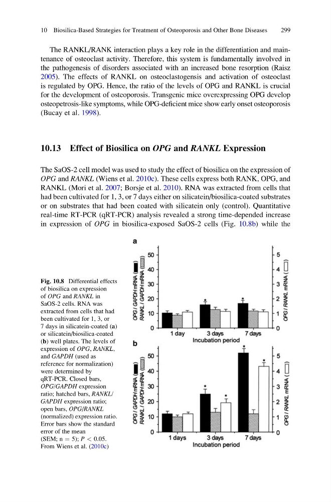
real-time RT-PCR (qRT-PCR) analysis revealed a strong time-depended increase
in expression of OPG in biosilica-exposed SaOS-2 cells (Fig. 10.8b) while the
Fig. 10.8 Differential effects
of biosilica on expression
of OPG and RANKL in
SaOS-2 cells. RNA was
extracted from cells that had
been cultivated for 1, 3, or
7 days in silicatein-coated (a)
or silicatein/biosilica-coated
(b) well plates. The levels of
expression of OPG, RANKL,
and GAPDH (used as
reference for normalization)
were determined by
qRT-PCR. Closed bars,
OPG/GAPDH expression
ratio; hatched bars, RANKL/
GAPDH expression ratio;
open bars, OPG/RANKL
(normalized) expression ratio.
Error bars show the standard
error of the mean
(SEM; n ¼ 5); P < 0.05.
From Wiens et al. (2010c)
10 Biosilica-Based Strategies for Treatment of Osteoporosis and Other Bone Diseases
299
the pathogenesis of disorders associated with an increased bone resorption (Raisz
2005). The effects of RANKL on osteoclastogensis and activation of osteoclast
is regulated by OPG. Hence, the ratio of the levels of OPG and RANKL is crucial
for the development of osteoporosis. Transgenic mice overexpressing OPG develop
osteopetrosis-like symptoms, while OPG-deficient mice show early onset osteoporosis
(Bucay et al. 1998).
10.13 Effect of Biosilica on OPG and RANKL Expression
The SaOS-2 cell model was used to study the effect of biosilica on the expression of
OPG and RANKL (Wiens et al. 2010c). These cells express both RANK, OPG, and
RANKL (Mori et al. 2007; Borsje et al. 2010). RNA was extracted from cells that
had been cultivated for 1, 3, or 7 days either on silicatein/biosilica-coated substrates
or on substrates that had been coated with silicatein only (control). Quantitative
real-time RT-PCR (qRT-PCR) analysis revealed a strong time-depended increase
in expression of OPG in biosilica-exposed SaOS-2 cells (Fig. 10.8b) while the
Fig. 10.8 Differential effects
of biosilica on expression
of OPG and RANKL in
SaOS-2 cells. RNA was
extracted from cells that had
been cultivated for 1, 3, or
7 days in silicatein-coated (a)
or silicatein/biosilica-coated
(b) well plates. The levels of
expression of OPG, RANKL,
and GAPDH (used as
reference for normalization)
were determined by
qRT-PCR. Closed bars,
OPG/GAPDH expression
ratio; hatched bars, RANKL/
GAPDH expression ratio;
open bars, OPG/RANKL
(normalized) expression ratio.
Error bars show the standard
error of the mean
(SEM; n ¼ 5); P < 0.05.
From Wiens et al. (2010c)
10 Biosilica-Based Strategies for Treatment of Osteoporosis and Other Bone Diseases
299