

10.10 Osteoinductive Index
A measure for the osteoinductive activity of a biomaterial is the osteoinductive index
which has been defined as the ratio between rate of [
3 H]dT incorporation into DNA
of bone (precursor) cells in vitro and the extent of bone (HA) formation in vivo
(Adkisson et al. 2000). To determine the osteoinductive activity in SaOS-2 cells, an
in vitro bioassay has been introduced, which is based on the calculation of the ratio
of two in vitro parameters, (1) cell proliferative activity, measured as the rate of [
3
H]
dT incorporation, and (2) biomineralization activity, measured by using the Alizarin
Red S staining assay of HA (Wiens et al. 2010b). SaOS-2 cells were incubated on
silicatein/biosilica-coated or uncoated substrates in medium containing ascorbic
acid and ß-glycerophosphate. Then, the cells were exposed to [
3
H]dT. Finally, the
incorporation of [
3
H]dT into DNA determined in a scintillation counter. Counts were
normalized to the total amount of DNA. Applying this assay, cultivation of SaOS2 cells on silicatein/biosilica-coated substrates was found to elicit a significant
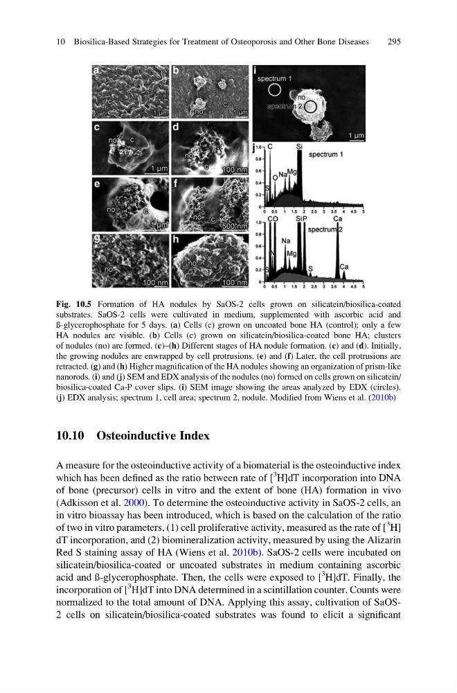
Fig. 10.5 Formation of HA nodules by SaOS-2 cells grown on silicatein/biosilica-coated
substrates. SaOS-2 cells were cultivated in medium, supplemented with ascorbic acid and
ß-glycerophosphate for 5 days. (a) Cells (c) grown on uncoated bone HA (control); only a few
HA nodules are visible. (b) Cells (c) grown on silicatein/biosilica-coated bone HA; clusters
of nodules (no) are formed. (c)–(h) Different stages of HA nodule formation. (c) and (d). Initially,
the growing nodules are enwrapped by cell protrusions. (e) and (f) Later, the cell protrusions are
retracted. (g) and (h) Higher magnification of the HA nodules showing an organization of prism-like
nanorods. (i) and (j) SEM and EDX analysis of the nodules (no) formed on cells grown on silicatein/
biosilica-coated Ca-P cover slips. (i) SEM image showing the areas analyzed by EDX (circles).
(j) EDX analysis; spectrum 1, cell area; spectrum 2, nodule. Modified from Wiens et al. (2010b)
10 Biosilica-Based Strategies for Treatment of Osteoporosis and Other Bone Diseases
295
A measure for the osteoinductive activity of a biomaterial is the osteoinductive index
which has been defined as the ratio between rate of [
3 H]dT incorporation into DNA
of bone (precursor) cells in vitro and the extent of bone (HA) formation in vivo
(Adkisson et al. 2000). To determine the osteoinductive activity in SaOS-2 cells, an
in vitro bioassay has been introduced, which is based on the calculation of the ratio
of two in vitro parameters, (1) cell proliferative activity, measured as the rate of [
3
H]
dT incorporation, and (2) biomineralization activity, measured by using the Alizarin
Red S staining assay of HA (Wiens et al. 2010b). SaOS-2 cells were incubated on
silicatein/biosilica-coated or uncoated substrates in medium containing ascorbic
acid and ß-glycerophosphate. Then, the cells were exposed to [
3
H]dT. Finally, the
incorporation of [
3
H]dT into DNA determined in a scintillation counter. Counts were
normalized to the total amount of DNA. Applying this assay, cultivation of SaOS2 cells on silicatein/biosilica-coated substrates was found to elicit a significant
Fig. 10.5 Formation of HA nodules by SaOS-2 cells grown on silicatein/biosilica-coated
substrates. SaOS-2 cells were cultivated in medium, supplemented with ascorbic acid and
ß-glycerophosphate for 5 days. (a) Cells (c) grown on uncoated bone HA (control); only a few
HA nodules are visible. (b) Cells (c) grown on silicatein/biosilica-coated bone HA; clusters
of nodules (no) are formed. (c)–(h) Different stages of HA nodule formation. (c) and (d). Initially,
the growing nodules are enwrapped by cell protrusions. (e) and (f) Later, the cell protrusions are
retracted. (g) and (h) Higher magnification of the HA nodules showing an organization of prism-like
nanorods. (i) and (j) SEM and EDX analysis of the nodules (no) formed on cells grown on silicatein/
biosilica-coated Ca-P cover slips. (i) SEM image showing the areas analyzed by EDX (circles).
(j) EDX analysis; spectrum 1, cell area; spectrum 2, nodule. Modified from Wiens et al. (2010b)
10 Biosilica-Based Strategies for Treatment of Osteoporosis and Other Bone Diseases
295