

crust to form together with oxygen silica and silica polymers. At present, the
mechanism by which sponges form the biosilica of their spicules is understood
in first outlines and has already enormously stimulated technologies in biomedicine (bone replacement or immuno-masking), in electronics (semiconductor
technologies), in optics (light transmission), in lithography, etc. With the application of the silicatein and the silicase enzymes, one could, in principle, envisage the
combination of three-dimensional architectures with silica structures to build threedimensional electronic biosilica chips.
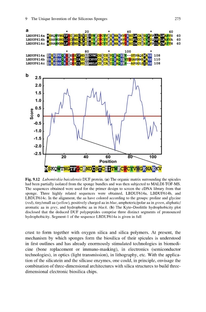
Fig. 9.12 Lubomirskia baicalensis DUF protein. (a) The organic matrix surrounding the spicules
had been partially isolated from the sponge bundles and was then subjected to MALDI-TOF-MS.
The sequences obtained were used for the primer design to screen the cDNA library from that
sponge. Three highly related sequences were obtained, LBDUF614a, LBDUF614b, and
LBDUF614c. In the alignment, the aa have colored according to the groups: proline and glycine
(red), tiny/small aa (yellow), positively charged aa in blue, amphoteric/polar aa in green, aliphatic/
aromatic aa in grey, and hydrophobic aa in black. (b) The Kyte–Doolittle hydrophobicity plot
disclosed that the deduced DUF polypeptides comprise three distinct segments of pronounced
hydrophobicity. Segment-1 of the sequence LBDUF614a is given in full
9 The Unique Invention of the Siliceous Sponges
275
mechanism by which sponges form the biosilica of their spicules is understood
in first outlines and has already enormously stimulated technologies in biomedicine (bone replacement or immuno-masking), in electronics (semiconductor
technologies), in optics (light transmission), in lithography, etc. With the application of the silicatein and the silicase enzymes, one could, in principle, envisage the
combination of three-dimensional architectures with silica structures to build threedimensional electronic biosilica chips.
Fig. 9.12 Lubomirskia baicalensis DUF protein. (a) The organic matrix surrounding the spicules
had been partially isolated from the sponge bundles and was then subjected to MALDI-TOF-MS.
The sequences obtained were used for the primer design to screen the cDNA library from that
sponge. Three highly related sequences were obtained, LBDUF614a, LBDUF614b, and
LBDUF614c. In the alignment, the aa have colored according to the groups: proline and glycine
(red), tiny/small aa (yellow), positively charged aa in blue, amphoteric/polar aa in green, aliphatic/
aromatic aa in grey, and hydrophobic aa in black. (b) The Kyte–Doolittle hydrophobicity plot
disclosed that the deduced DUF polypeptides comprise three distinct segments of pronounced
hydrophobicity. Segment-1 of the sequence LBDUF614a is given in full
9 The Unique Invention of the Siliceous Sponges
275