

Based on these microscopic data, first clues on the factors involved in morphogenesis of the spicules could be outlined. It is obvious that the size/length of the
spicules (megascleres), size >50 mm, exceeds that of a cell. Therefore, several
mechanisms might be postulated for the intracellular and subsequent extracellular
growth of the spicules, giving rise to a complex morphology. Sollas (1888) assumed
that cells migrate along the spicules to allow growth along their axis. This view was
strongly supported by Maas (1901). Studying the demosponge Tethya lyncurium, he
observed (what was forgotten later) that initially the synthesis of the spicules starts
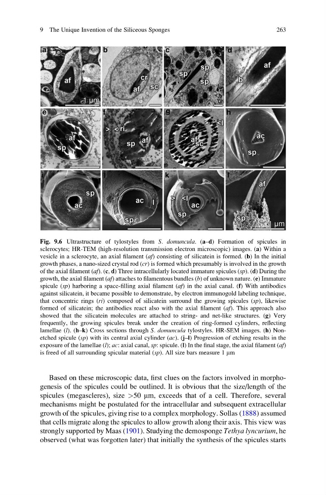
Fig. 9.6 Ultrastructure of tylostyles from S. domuncula. (a–d) Formation of spicules in
sclerocytes; HR-TEM (high-resolution transmission electron microscopic) images. (a) Within a
vesicle in a sclerocyte, an axial filament (af) consisting of silicatein is formed. (b) In the initial
growth phases, a nano-sized crystal rod (cr) is formed which presumably is involved in the growth
of the axial filament (af). (c, d) Three intracellularly located immature spicules (sp). (d) During the
growth, the axial filament (af) attaches to filamentous bundles (b) of unknown nature. (e) Immature
spicule (sp) harboring a space-filling axial filament (af) in the axial canal. (f) With antibodies
against silicatein, it became possible to demonstrate, by electron immunogold labeling technique,
that concentric rings (ri) composed of silicatein surround the growing spicules (sp), likewise
formed of silicatein; the antibodies react also with the axial filament (af). This approach also
showed that the silicatein molecules are attached to string- and net-like structures. (g) Very
frequently, the growing spicules break under the creation of ring-formed cylinders, reflecting
lamellae (l). (h–k) Cross sections through S. domuncula tylostyles. HR-SEM images. (h) Nonetched spicule (sp) with its central axial cylinder (ac). (j–l) Progression of etching results in the
exposure of the lamellae (l); ac: axial canal, sp: spicule. (l) In the final stage, the axial filament (af)
is freed of all surrounding spicular material (sp). All size bars measure 1 mm
9 The Unique Invention of the Siliceous Sponges
263
spicules (megascleres), size >50 mm, exceeds that of a cell. Therefore, several
mechanisms might be postulated for the intracellular and subsequent extracellular
growth of the spicules, giving rise to a complex morphology. Sollas (1888) assumed
that cells migrate along the spicules to allow growth along their axis. This view was
strongly supported by Maas (1901). Studying the demosponge Tethya lyncurium, he
observed (what was forgotten later) that initially the synthesis of the spicules starts
Fig. 9.6 Ultrastructure of tylostyles from S. domuncula. (a–d) Formation of spicules in
sclerocytes; HR-TEM (high-resolution transmission electron microscopic) images. (a) Within a
vesicle in a sclerocyte, an axial filament (af) consisting of silicatein is formed. (b) In the initial
growth phases, a nano-sized crystal rod (cr) is formed which presumably is involved in the growth
of the axial filament (af). (c, d) Three intracellularly located immature spicules (sp). (d) During the
growth, the axial filament (af) attaches to filamentous bundles (b) of unknown nature. (e) Immature
spicule (sp) harboring a space-filling axial filament (af) in the axial canal. (f) With antibodies
against silicatein, it became possible to demonstrate, by electron immunogold labeling technique,
that concentric rings (ri) composed of silicatein surround the growing spicules (sp), likewise
formed of silicatein; the antibodies react also with the axial filament (af). This approach also
showed that the silicatein molecules are attached to string- and net-like structures. (g) Very
frequently, the growing spicules break under the creation of ring-formed cylinders, reflecting
lamellae (l). (h–k) Cross sections through S. domuncula tylostyles. HR-SEM images. (h) Nonetched spicule (sp) with its central axial cylinder (ac). (j–l) Progression of etching results in the
exposure of the lamellae (l); ac: axial canal, sp: spicule. (l) In the final stage, the axial filament (af)
is freed of all surrounding spicular material (sp). All size bars measure 1 mm
9 The Unique Invention of the Siliceous Sponges
263