

2005). The immune serum showed a dense accumulation of gold granules in the
sclerocytes and the extracellular space. Fine structure analysis revealed that at first
concentric rings which are 0.2–0.5 mm apart are seen around the forming spicules
(Fig. 9.6f, g). Subsequently, the inner rings fuse and electron-dense clouds become
visible. Later, during maturation, the number of the concentric rings increases from
two to ten rings with a total diameter 4–6 mm. These data underscore the view that
the spicules grow appositionally in both directions, in width and in length (reviewed
in M€ uller et al. 2006b, 2007c, 2009). Limited etching of the spicules with
hydrofluoric acid displays that the initially formed and separately existing lamellae
can still be distinguished (Fig. 9.6i–k). Finally, the spicules release the axial
filament from the bio-silica mantel (Fig. 9.6l).
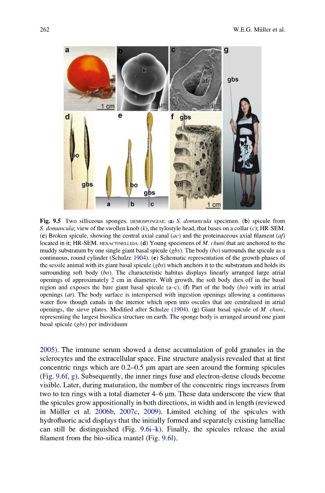
Fig. 9.5 Two silliceous sponges. DEMOSPONGIAE: (a) S. domuncula specimen. (b) spicule from
S. domuncula; view of the swollen knob (k), the tylostyle head, that bases on a collar (c); HR-SEM.
(c) Broken spicule, showing the central axial canal (ac) and the proteinaceous axial filament (af)
located in it; HR-SEM. HEXACTINELLIDA: (d) Young specimens of M. chuni that are anchored to the
muddy substratum by one single giant basal spicule (gbs). The body (bo) surrounds the spicule as a
continuous, round cylinder (Schulze 1904). (e) Schematic representation of the growth phases of
the sessile animal with its giant basal spicule (gbs) which anchors it to the substratum and holds its
surrounding soft body (bo). The characteristic habitus displays linearly arranged large atrial
openings of approximately 2 cm in diameter. With growth, the soft body dies off in the basal
region and exposes the bare giant basal spicule (a–c). (f) Part of the body (bo) with its atrial
openings (at). The body surface is interspersed with ingestion openings allowing a continuous
water flow though canals in the interior which open into oscules that are centralized in atrial
openings, the sieve plates. Modified after Schulze (1904). (g) Giant basal spicule of M. chuni,
representing the largest biosilica structure on earth. The sponge body is arranged around one giant
basal spicule (gbs) per individuum
262
W.E.G. M€ uller et al.
sclerocytes and the extracellular space. Fine structure analysis revealed that at first
concentric rings which are 0.2–0.5 mm apart are seen around the forming spicules
(Fig. 9.6f, g). Subsequently, the inner rings fuse and electron-dense clouds become
visible. Later, during maturation, the number of the concentric rings increases from
two to ten rings with a total diameter 4–6 mm. These data underscore the view that
the spicules grow appositionally in both directions, in width and in length (reviewed
in M€ uller et al. 2006b, 2007c, 2009). Limited etching of the spicules with
hydrofluoric acid displays that the initially formed and separately existing lamellae
can still be distinguished (Fig. 9.6i–k). Finally, the spicules release the axial
filament from the bio-silica mantel (Fig. 9.6l).
Fig. 9.5 Two silliceous sponges. DEMOSPONGIAE: (a) S. domuncula specimen. (b) spicule from
S. domuncula; view of the swollen knob (k), the tylostyle head, that bases on a collar (c); HR-SEM.
(c) Broken spicule, showing the central axial canal (ac) and the proteinaceous axial filament (af)
located in it; HR-SEM. HEXACTINELLIDA: (d) Young specimens of M. chuni that are anchored to the
muddy substratum by one single giant basal spicule (gbs). The body (bo) surrounds the spicule as a
continuous, round cylinder (Schulze 1904). (e) Schematic representation of the growth phases of
the sessile animal with its giant basal spicule (gbs) which anchors it to the substratum and holds its
surrounding soft body (bo). The characteristic habitus displays linearly arranged large atrial
openings of approximately 2 cm in diameter. With growth, the soft body dies off in the basal
region and exposes the bare giant basal spicule (a–c). (f) Part of the body (bo) with its atrial
openings (at). The body surface is interspersed with ingestion openings allowing a continuous
water flow though canals in the interior which open into oscules that are centralized in atrial
openings, the sieve plates. Modified after Schulze (1904). (g) Giant basal spicule of M. chuni,
representing the largest biosilica structure on earth. The sponge body is arranged around one giant
basal spicule (gbs) per individuum
262
W.E.G. M€ uller et al.