

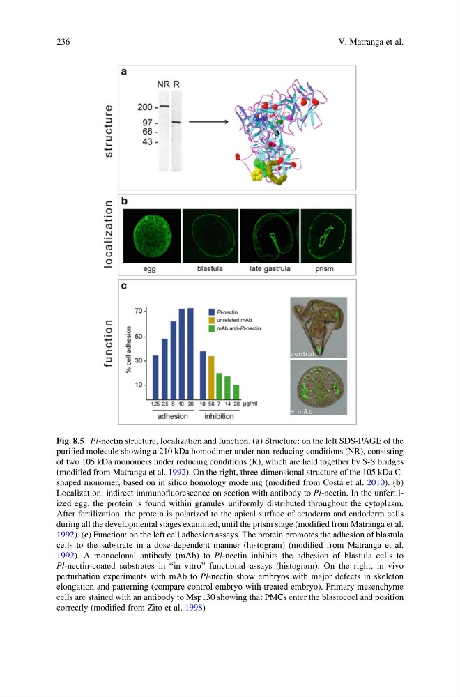
Fig. 8.5 Pl-nectin structure, localization and function. (a) Structure: on the left SDS-PAGE of the
purified molecule showing a 210 kDa homodimer under non-reducing conditions (NR), consisting
of two 105 kDa monomers under reducing conditions (R), which are held together by S-S bridges
(modified from Matranga et al. 1992). On the right, three-dimensional structure of the 105 kDa Cshaped monomer, based on in silico homology modeling (modified from Costa et al. 2010). (b)
Localization: indirect immunofluorescence on section with antibody to Pl-nectin. In the unfertilized egg, the protein is found within granules uniformly distributed throughout the cytoplasm.
After fertilization, the protein is polarized to the apical surface of ectoderm and endoderm cells
during all the developmental stages examined, until the prism stage (modified from Matranga et al.
1992). (c) Function: on the left cell adhesion assays. The protein promotes the adhesion of blastula
cells to the substrate in a dose-dependent manner (histogram) (modified from Matranga et al.
1992). A monoclonal antibody (mAb) to Pl-nectin inhibits the adhesion of blastula cells to
Pl-nectin-coated substrates in “in vitro” functional assays (histogram). On the right, in vivo
perturbation experiments with mAb to Pl-nectin show embryos with major defects in skeleton
elongation and patterning (compare control embryo with treated embryo). Primary mesenchyme
cells are stained with an antibody to Msp130 showing that PMCs enter the blastocoel and position
correctly (modified from Zito et al. 1998)
236
V. Matranga et al.
purified molecule showing a 210 kDa homodimer under non-reducing conditions (NR), consisting
of two 105 kDa monomers under reducing conditions (R), which are held together by S-S bridges
(modified from Matranga et al. 1992). On the right, three-dimensional structure of the 105 kDa Cshaped monomer, based on in silico homology modeling (modified from Costa et al. 2010). (b)
Localization: indirect immunofluorescence on section with antibody to Pl-nectin. In the unfertilized egg, the protein is found within granules uniformly distributed throughout the cytoplasm.
After fertilization, the protein is polarized to the apical surface of ectoderm and endoderm cells
during all the developmental stages examined, until the prism stage (modified from Matranga et al.
1992). (c) Function: on the left cell adhesion assays. The protein promotes the adhesion of blastula
cells to the substrate in a dose-dependent manner (histogram) (modified from Matranga et al.
1992). A monoclonal antibody (mAb) to Pl-nectin inhibits the adhesion of blastula cells to
Pl-nectin-coated substrates in “in vitro” functional assays (histogram). On the right, in vivo
perturbation experiments with mAb to Pl-nectin show embryos with major defects in skeleton
elongation and patterning (compare control embryo with treated embryo). Primary mesenchyme
cells are stained with an antibody to Msp130 showing that PMCs enter the blastocoel and position
correctly (modified from Zito et al. 1998)
236
V. Matranga et al.