

information to migrating PMCs. Interestingly, ECM3 contains an N-terminal
domain similar to the mammalian chondroitin sulfate proteoglycan core protein
NG2, required for the responsiveness of some cell types to PDGF, supporting the
idea that it may play a role in growth factor presentation (Hodor et al. 2000). The
central region of ECM3 contains five tandem repeats of a motif similar to the
domain contained within the regulatory Ca
2+- binding loop of the Na
+ -Ca
2+
exchange protein, which is similar in several respects to MAFp4, a component of
the aggregation factor molecular complex, mediating the species-specific sorting of
sponge cells (Hodor et al. 2000).
Other ECM proteins located on the basal lamina have been described to have a
role in spicule formation. For example, by microinjection of specific antibodies into
the blastocoelic cavity, it has been shown that collagen in S. purpuratus (Wessel
et al. 1991) and Pl-200 K protein in P. lividus (Tesoro et al. 1998) are essential for
skeleton elongation and patterning.
Sea urchin metalloproteinases can modify collagen and other ECM components,
as well as glycoproteins that associate with collagens and carbohydrates in the
ECM. It has been shown that several inhibitors of metalloproteinases inhibit the
continuation of skeletogenesis in both S. purpuratus and L. pictus embryos
(Ingersoll and Wilt 1998).
The functional role played by ECM molecules in vivo has often been
investigated by means of monoclonal antibodies (McAb) which interfere with
their functions. Among the ECM proteins already known, Pl-nectin, isolated from
P. lividus (Matranga et al. 1992) as a collagen-binding molecule, is the first
described as an “indirect actor” in the ecto-mesoderm signaling (Zito et al. 1998).
The protein, a discoidin family member whose complete sequence and domain
architecture has been recently characterized (Fig. 8.5a) (Costa et al. 2010), is
localized on the apical surface of ectoderm and endoderm cells from the blastula
and gastrula stage onwards (Fig. 8.5b). By in vitro assays, it has been shown to
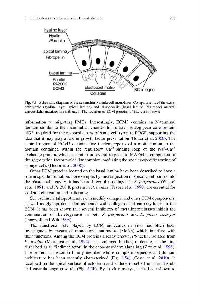
Fig. 8.4 Schematic diagram of the sea urchin blastula cell monolayer. Compartments of the extraembryonic (hyaline layer, apical lamina) and blastocoelic (basal lamina, blastocoel matrix)
extracellular matrixes are indicated. The location of ECM proteins of interest is shown
8 Echinoderms as Blueprints for Biocalcification
235
domain similar to the mammalian chondroitin sulfate proteoglycan core protein
NG2, required for the responsiveness of some cell types to PDGF, supporting the
idea that it may play a role in growth factor presentation (Hodor et al. 2000). The
central region of ECM3 contains five tandem repeats of a motif similar to the
domain contained within the regulatory Ca
2+- binding loop of the Na
+ -Ca
2+
exchange protein, which is similar in several respects to MAFp4, a component of
the aggregation factor molecular complex, mediating the species-specific sorting of
sponge cells (Hodor et al. 2000).
Other ECM proteins located on the basal lamina have been described to have a
role in spicule formation. For example, by microinjection of specific antibodies into
the blastocoelic cavity, it has been shown that collagen in S. purpuratus (Wessel
et al. 1991) and Pl-200 K protein in P. lividus (Tesoro et al. 1998) are essential for
skeleton elongation and patterning.
Sea urchin metalloproteinases can modify collagen and other ECM components,
as well as glycoproteins that associate with collagens and carbohydrates in the
ECM. It has been shown that several inhibitors of metalloproteinases inhibit the
continuation of skeletogenesis in both S. purpuratus and L. pictus embryos
(Ingersoll and Wilt 1998).
The functional role played by ECM molecules in vivo has often been
investigated by means of monoclonal antibodies (McAb) which interfere with
their functions. Among the ECM proteins already known, Pl-nectin, isolated from
P. lividus (Matranga et al. 1992) as a collagen-binding molecule, is the first
described as an “indirect actor” in the ecto-mesoderm signaling (Zito et al. 1998).
The protein, a discoidin family member whose complete sequence and domain
architecture has been recently characterized (Fig. 8.5a) (Costa et al. 2010), is
localized on the apical surface of ectoderm and endoderm cells from the blastula
and gastrula stage onwards (Fig. 8.5b). By in vitro assays, it has been shown to
Fig. 8.4 Schematic diagram of the sea urchin blastula cell monolayer. Compartments of the extraembryonic (hyaline layer, apical lamina) and blastocoelic (basal lamina, blastocoel matrix)
extracellular matrixes are indicated. The location of ECM proteins of interest is shown
8 Echinoderms as Blueprints for Biocalcification
235