

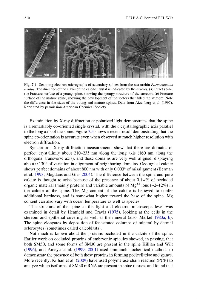
Examination by X-ray diffraction or polarized light demonstrates that the spine
is a remarkably co-oriented single crystal, with the c crystallographic axis parallel
to the long axis of the spine. Figure 7.5 shows a recent result demonstrating that the
spine co-orientation is accurate even when observed at much higher resolution with
electron diffraction.
Synchrotron X-ray diffraction measurements show that there are domains of
perfect crystallinity about 210–235 nm along the long axis (160 nm along the
orthogonal transverse axis), and these domains are very well aligned, displaying
about 0.130
of variation in alignment of neighboring domains. Geological calcite
shows perfect domains of about 800 nm with only 0.003
of misalignment (Berman
et al. 1993; Magdans and Gies 2004). The difference between the spine and pure
calcite is thought to arise because of the presence of about 0.1w% of occluded
organic material (mainly protein) and variable amounts of Mg
+2 ions (~2–12%) in
the calcite of the spine. The Mg content of the calcite is believed to confer
additional hardness, and is somewhat higher toward the base of the spine. Mg
content can also vary with ocean temperature as well as species.
The structure of the spine at the light and electron microscope level was
examined in detail by Heatfield and Travis (1975), looking at the cells in the
stereom and epithelial covering as well as the mineral (also, M€ arkel 1983a, b).
The spine elongates by deposition of fenestrated columns of mineral by dermal
sclerocytes (sometimes called calcoblasts).
Not much is known about the proteins occluded in the calcite of the spine.
Earlier work on occluded proteins of embryonic spicules showed, in passing, that
both SM50, and some forms of SM30 are present in the spine Killian and Wilt
(1996), and Ameye et al. (1999, 2001) used immunohistochemical methods to
demonstrate the presence of both these proteins in forming pedicellariae and spines.
More recently, Killian et al. (2009) have used polymerase chain reaction (PCR) to
analyze which isoforms of SM30 mRNA are present in spine tissues, and found that
Fig. 7.4 Scanning electron micrographs of secondary spines from the sea urchin Paracentrotus
lividus. The direction of the c axis of the calcite crystal is indicated by the arrows. (a) Intact spine.
(b) Fracture surface of a young spine, showing the spongy structure of the stereom. (c) Fracture
surface of the mature spine, showing the development of the sectors that filled the stereom. Note
the difference in the sizes of the young and mature spines. Data from Aizenberg et al. (1997).
Reprinted by permission American Chemical Society
210
P.U.P.A Gilbert and F.H. Wilt
is a remarkably co-oriented single crystal, with the c crystallographic axis parallel
to the long axis of the spine. Figure 7.5 shows a recent result demonstrating that the
spine co-orientation is accurate even when observed at much higher resolution with
electron diffraction.
Synchrotron X-ray diffraction measurements show that there are domains of
perfect crystallinity about 210–235 nm along the long axis (160 nm along the
orthogonal transverse axis), and these domains are very well aligned, displaying
about 0.130
of variation in alignment of neighboring domains. Geological calcite
shows perfect domains of about 800 nm with only 0.003
of misalignment (Berman
et al. 1993; Magdans and Gies 2004). The difference between the spine and pure
calcite is thought to arise because of the presence of about 0.1w% of occluded
organic material (mainly protein) and variable amounts of Mg
+2 ions (~2–12%) in
the calcite of the spine. The Mg content of the calcite is believed to confer
additional hardness, and is somewhat higher toward the base of the spine. Mg
content can also vary with ocean temperature as well as species.
The structure of the spine at the light and electron microscope level was
examined in detail by Heatfield and Travis (1975), looking at the cells in the
stereom and epithelial covering as well as the mineral (also, M€ arkel 1983a, b).
The spine elongates by deposition of fenestrated columns of mineral by dermal
sclerocytes (sometimes called calcoblasts).
Not much is known about the proteins occluded in the calcite of the spine.
Earlier work on occluded proteins of embryonic spicules showed, in passing, that
both SM50, and some forms of SM30 are present in the spine Killian and Wilt
(1996), and Ameye et al. (1999, 2001) used immunohistochemical methods to
demonstrate the presence of both these proteins in forming pedicellariae and spines.
More recently, Killian et al. (2009) have used polymerase chain reaction (PCR) to
analyze which isoforms of SM30 mRNA are present in spine tissues, and found that
Fig. 7.4 Scanning electron micrographs of secondary spines from the sea urchin Paracentrotus
lividus. The direction of the c axis of the calcite crystal is indicated by the arrows. (a) Intact spine.
(b) Fracture surface of a young spine, showing the spongy structure of the stereom. (c) Fracture
surface of the mature spine, showing the development of the sectors that filled the stereom. Note
the difference in the sizes of the young and mature spines. Data from Aizenberg et al. (1997).
Reprinted by permission American Chemical Society
210
P.U.P.A Gilbert and F.H. Wilt