

descendants, which are now called primary mesenchyme cells (PMC), then form a
stereotypical array in the vegetal portion of the blastocoel, and adjacent PMCs fuse
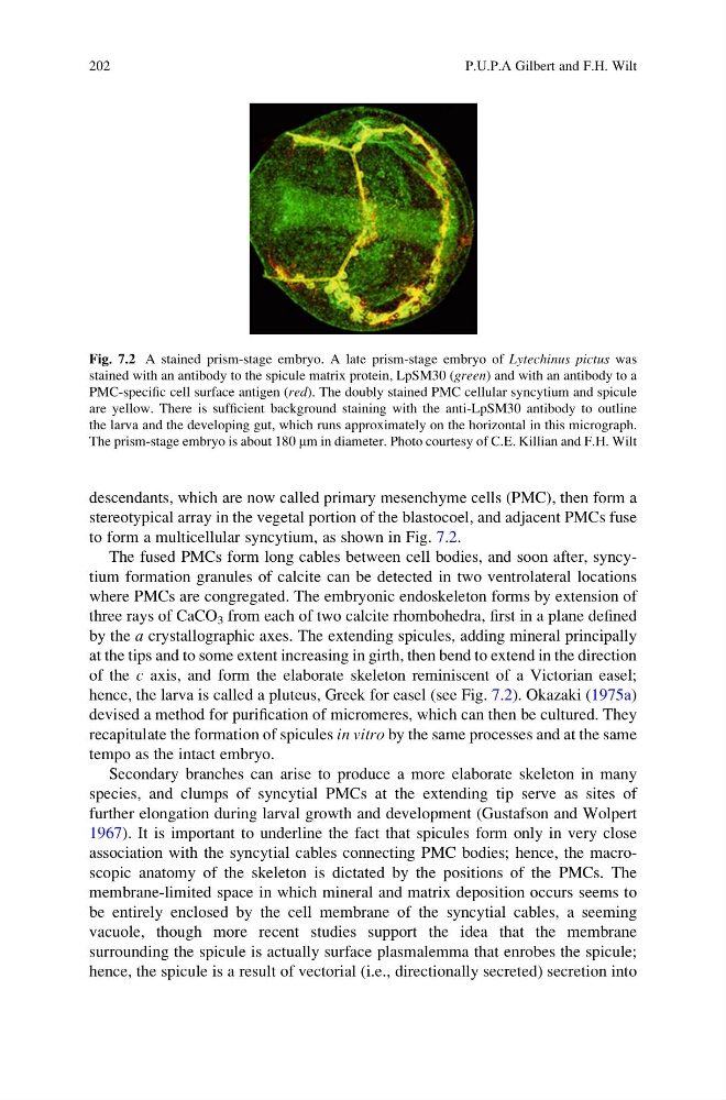
to form a multicellular syncytium, as shown in Fig. 7.2.
The fused PMCs form long cables between cell bodies, and soon after, syncytium formation granules of calcite can be detected in two ventrolateral locations
where PMCs are congregated. The embryonic endoskeleton forms by extension of
three rays of CaCO 3 from each of two calcite rhombohedra, first in a plane defined
by the a crystallographic axes. The extending spicules, adding mineral principally
at the tips and to some extent increasing in girth, then bend to extend in the direction
of the c axis, and form the elaborate skeleton reminiscent of a Victorian easel;
hence, the larva is called a pluteus, Greek for easel (see Fig. 7.2). Okazaki (1975a)
devised a method for purification of micromeres, which can then be cultured. They
recapitulate the formation of spicules in vitro by the same processes and at the same
tempo as the intact embryo.
Secondary branches can arise to produce a more elaborate skeleton in many
species, and clumps of syncytial PMCs at the extending tip serve as sites of
further elongation during larval growth and development (Gustafson and Wolpert
1967). It is important to underline the fact that spicules form only in very close
association with the syncytial cables connecting PMC bodies; hence, the macroscopic anatomy of the skeleton is dictated by the positions of the PMCs. The
membrane-limited space in which mineral and matrix deposition occurs seems to
be entirely enclosed by the cell membrane of the syncytial cables, a seeming
vacuole, though more recent studies support the idea that the membrane
surrounding the spicule is actually surface plasmalemma that enrobes the spicule;
hence, the spicule is a result of vectorial (i.e., directionally secreted) secretion into
Fig. 7.2 A stained prism-stage embryo. A late prism-stage embryo of Lytechinus pictus was
stained with an antibody to the spicule matrix protein, LpSM30 (green) and with an antibody to a
PMC-specific cell surface antigen (red). The doubly stained PMC cellular syncytium and spicule
are yellow. There is sufficient background staining with the anti-LpSM30 antibody to outline
the larva and the developing gut, which runs approximately on the horizontal in this micrograph.
The prism-stage embryo is about 180 mm in diameter. Photo courtesy of C.E. Killian and F.H. Wilt
202
P.U.P.A Gilbert and F.H. Wilt
stereotypical array in the vegetal portion of the blastocoel, and adjacent PMCs fuse
to form a multicellular syncytium, as shown in Fig. 7.2.
The fused PMCs form long cables between cell bodies, and soon after, syncytium formation granules of calcite can be detected in two ventrolateral locations
where PMCs are congregated. The embryonic endoskeleton forms by extension of
three rays of CaCO 3 from each of two calcite rhombohedra, first in a plane defined
by the a crystallographic axes. The extending spicules, adding mineral principally
at the tips and to some extent increasing in girth, then bend to extend in the direction
of the c axis, and form the elaborate skeleton reminiscent of a Victorian easel;
hence, the larva is called a pluteus, Greek for easel (see Fig. 7.2). Okazaki (1975a)
devised a method for purification of micromeres, which can then be cultured. They
recapitulate the formation of spicules in vitro by the same processes and at the same
tempo as the intact embryo.
Secondary branches can arise to produce a more elaborate skeleton in many
species, and clumps of syncytial PMCs at the extending tip serve as sites of
further elongation during larval growth and development (Gustafson and Wolpert
1967). It is important to underline the fact that spicules form only in very close
association with the syncytial cables connecting PMC bodies; hence, the macroscopic anatomy of the skeleton is dictated by the positions of the PMCs. The
membrane-limited space in which mineral and matrix deposition occurs seems to
be entirely enclosed by the cell membrane of the syncytial cables, a seeming
vacuole, though more recent studies support the idea that the membrane
surrounding the spicule is actually surface plasmalemma that enrobes the spicule;
hence, the spicule is a result of vectorial (i.e., directionally secreted) secretion into
Fig. 7.2 A stained prism-stage embryo. A late prism-stage embryo of Lytechinus pictus was
stained with an antibody to the spicule matrix protein, LpSM30 (green) and with an antibody to a
PMC-specific cell surface antigen (red). The doubly stained PMC cellular syncytium and spicule
are yellow. There is sufficient background staining with the anti-LpSM30 antibody to outline
the larva and the developing gut, which runs approximately on the horizontal in this micrograph.
The prism-stage embryo is about 180 mm in diameter. Photo courtesy of C.E. Killian and F.H. Wilt
202
P.U.P.A Gilbert and F.H. Wilt