

7.2 Formation of the Endoskeleton in the Embryo
7.2.1 Spicule Formation
The development of the larval endoskeleton in indirectly developing sea urchins
(i.e., those that develop through a larval stage) has been studied extensively by
embryologists since the late 1800s. The formation of the larval endoskeleton can
be followed in real time under the microscope. At the dawning of modern experimental biology, the endoskeletal spicules found in sea urchin larvae were the
morphological characters Boveri used in his discovery that chromosomes were
the carriers of heredity (Laubichler and Davidson 2008).
The embryology of spicule development has been described in detail by Okazaki
(1975a, b), and more recently by Wilt and Ettensohn (2007). At the fourth cell
division, four of the resultant 16 cells are clustered at one pole, and are termed
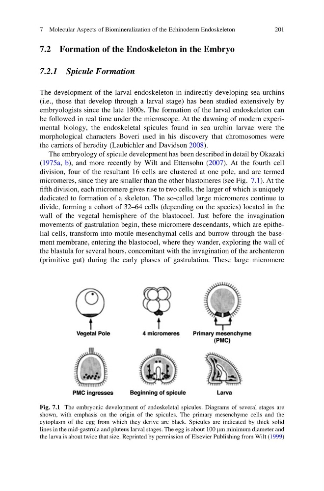
micromeres, since they are smaller than the other blastomeres (see Fig. 7.1). At the
fifth division, each micromere gives rise to two cells, the larger of which is uniquely
dedicated to formation of a skeleton. The so-called large micromeres continue to
divide, forming a cohort of 32–64 cells (depending on the species) located in the
wall of the vegetal hemisphere of the blastocoel. Just before the invagination
movements of gastrulation begin, these micromere descendants, which are epithelial cells, transform into motile mesenchymal cells and burrow through the basement membrane, entering the blastocoel, where they wander, exploring the wall of
the blastula for several hours, concomitant with the invagination of the archenteron
(primitive gut) during the early phases of gastrulation. These large micromere
Fig. 7.1 The embryonic development of endoskeletal spicules. Diagrams of several stages are
shown, with emphasis on the origin of the spicules. The primary mesenchyme cells and the
cytoplasm of the egg from which they derive are black. Spicules are indicated by thick solid
lines in the mid-gastrula and pluteus larval stages. The egg is about 100 mm minimum diameter and
the larva is about twice that size. Reprinted by permission of Elsevier Publishing from Wilt (1999)
7 Molecular Aspects of Biomineralization of the Echinoderm Endoskeleton
201
7.2.1 Spicule Formation
The development of the larval endoskeleton in indirectly developing sea urchins
(i.e., those that develop through a larval stage) has been studied extensively by
embryologists since the late 1800s. The formation of the larval endoskeleton can
be followed in real time under the microscope. At the dawning of modern experimental biology, the endoskeletal spicules found in sea urchin larvae were the
morphological characters Boveri used in his discovery that chromosomes were
the carriers of heredity (Laubichler and Davidson 2008).
The embryology of spicule development has been described in detail by Okazaki
(1975a, b), and more recently by Wilt and Ettensohn (2007). At the fourth cell
division, four of the resultant 16 cells are clustered at one pole, and are termed
micromeres, since they are smaller than the other blastomeres (see Fig. 7.1). At the
fifth division, each micromere gives rise to two cells, the larger of which is uniquely
dedicated to formation of a skeleton. The so-called large micromeres continue to
divide, forming a cohort of 32–64 cells (depending on the species) located in the
wall of the vegetal hemisphere of the blastocoel. Just before the invagination
movements of gastrulation begin, these micromere descendants, which are epithelial cells, transform into motile mesenchymal cells and burrow through the basement membrane, entering the blastocoel, where they wander, exploring the wall of
the blastula for several hours, concomitant with the invagination of the archenteron
(primitive gut) during the early phases of gastrulation. These large micromere
Fig. 7.1 The embryonic development of endoskeletal spicules. Diagrams of several stages are
shown, with emphasis on the origin of the spicules. The primary mesenchyme cells and the
cytoplasm of the egg from which they derive are black. Spicules are indicated by thick solid
lines in the mid-gastrula and pluteus larval stages. The egg is about 100 mm minimum diameter and
the larva is about twice that size. Reprinted by permission of Elsevier Publishing from Wilt (1999)
7 Molecular Aspects of Biomineralization of the Echinoderm Endoskeleton
201