

the relative orientation of the HA crystals with respect to collagen fibrils, electron
diffraction investigation have been carried out. The results demonstrate the preferential alignment of the HA crystallographic c-axis with the collagen fibril longitudinal axis. To study the relationship between the newly formed crystals and the
collagen fibrils directly, high-resolution TEM (HRTEM) analysis of the lattice
plane arrangements was performed (Fig. 6.29b). The HRTEM analysis of the
parallel-aligned mineralized collagen fibrils revealed that crystal lattice is seen
not only on the side area of the collagen fibrils, but also in the middle area, and
that the electron density on the surface of the collagen fibrils is higher than in the
interior area. These findings indicated that HA crystals grew on the surface of the
collagen fibrils, giving the first direct evidence to support previous theories (Cui
et al. 2007).
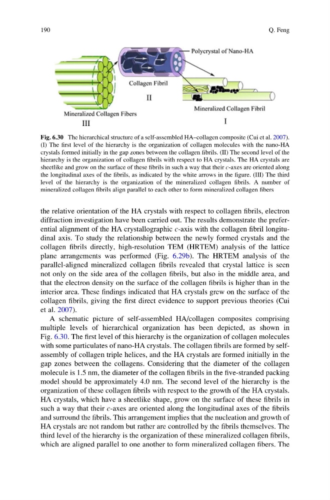
A schematic picture of self-assembled HA/collagen composites comprising
multiple levels of hierarchical organization has been depicted, as shown in
Fig. 6.30. The first level of this hierarchy is the organization of collagen molecules
with some particulates of nano-HA crystals. The collagen fibrils are formed by selfassembly of collagen triple helices, and the HA crystals are formed initially in the
gap zones between the collagens. Considering that the diameter of the collagen
molecule is 1.5 nm, the diameter of the collagen fibrils in the five-stranded packing
model should be approximately 4.0 nm. The second level of the hierarchy is the
organization of these collagen fibrils with respect to the growth of the HA crystals.
HA crystals, which have a sheetlike shape, grow on the surface of these fibrils in
such a way that their c-axes are oriented along the longitudinal axes of the fibrils
and surround the fibrils. This arrangement implies that the nucleation and growth of
HA crystals are not random but rather are controlled by the fibrils themselves. The
third level of the hierarchy is the organization of these mineralized collagen fibrils,
which are aligned parallel to one another to form mineralized collagen fibers. The
Fig. 6.30 The hierarchical structure of a self-assembled HA–collagen composite (Cui et al. 2007).
(I) The first level of the hierarchy is the organization of collagen molecules with the nano-HA
crystals formed initially in the gap zones between the collagen fibrils. (II) The second level of the
hierarchy is the organization of collagen fibrils with respect to HA crystals. The HA crystals are
sheetlike and grow on the surface of these fibrils in such a way that their c-axes are oriented along
the longitudinal axes of the fibrils, as indicated by the white arrows in the figure. (III) The third
level of the hierarchy is the organization of the mineralized collagen fibrils. A number of
mineralized collagen fibrils align parallel to each other to form mineralized collagen fibers
190
Q. Feng
diffraction investigation have been carried out. The results demonstrate the preferential alignment of the HA crystallographic c-axis with the collagen fibril longitudinal axis. To study the relationship between the newly formed crystals and the
collagen fibrils directly, high-resolution TEM (HRTEM) analysis of the lattice
plane arrangements was performed (Fig. 6.29b). The HRTEM analysis of the
parallel-aligned mineralized collagen fibrils revealed that crystal lattice is seen
not only on the side area of the collagen fibrils, but also in the middle area, and
that the electron density on the surface of the collagen fibrils is higher than in the
interior area. These findings indicated that HA crystals grew on the surface of the
collagen fibrils, giving the first direct evidence to support previous theories (Cui
et al. 2007).
A schematic picture of self-assembled HA/collagen composites comprising
multiple levels of hierarchical organization has been depicted, as shown in
Fig. 6.30. The first level of this hierarchy is the organization of collagen molecules
with some particulates of nano-HA crystals. The collagen fibrils are formed by selfassembly of collagen triple helices, and the HA crystals are formed initially in the
gap zones between the collagens. Considering that the diameter of the collagen
molecule is 1.5 nm, the diameter of the collagen fibrils in the five-stranded packing
model should be approximately 4.0 nm. The second level of the hierarchy is the
organization of these collagen fibrils with respect to the growth of the HA crystals.
HA crystals, which have a sheetlike shape, grow on the surface of these fibrils in
such a way that their c-axes are oriented along the longitudinal axes of the fibrils
and surround the fibrils. This arrangement implies that the nucleation and growth of
HA crystals are not random but rather are controlled by the fibrils themselves. The
third level of the hierarchy is the organization of these mineralized collagen fibrils,
which are aligned parallel to one another to form mineralized collagen fibers. The
Fig. 6.30 The hierarchical structure of a self-assembled HA–collagen composite (Cui et al. 2007).
(I) The first level of the hierarchy is the organization of collagen molecules with the nano-HA
crystals formed initially in the gap zones between the collagen fibrils. (II) The second level of the
hierarchy is the organization of collagen fibrils with respect to HA crystals. The HA crystals are
sheetlike and grow on the surface of these fibrils in such a way that their c-axes are oriented along
the longitudinal axes of the fibrils, as indicated by the white arrows in the figure. (III) The third
level of the hierarchy is the organization of the mineralized collagen fibrils. A number of
mineralized collagen fibrils align parallel to each other to form mineralized collagen fibers
190
Q. Feng