

improve the attachment between the collagen fibrils and the calcium phosphate
crystals. Rhee et al. (2000) investigated the nucleation of Ca–P crystal through
chemical interaction with collagen by soaking a collagen membrane in a supersaturated simulated body fluid solution. Goissis et al. (2003) reported both in vitro and
in vivo studies of biomimetic mineralization of charged collagen matrices. Their
results showed that the calcium phosphate deposited in close resemblance to the
D-periodicity seen for assembly of collagen fibrils in vivo. Additionally, the in vitro
results suggested that amide hydrolysis may have introduced into the matrix, signs
for the controlled mineralization of collagen fiber. Amide hydrolysis was found
from TEM investigations to occur near the overlap and gap zones. Pederson et al.
(2003) reported a strategy for exploiting temperature-driven self-assembly of
collagen and thermally triggered liposome mineralization to form a mineralized
collagen composite. Cui et al. have now synthetically prepared nanofibrils of
mineralized collagen as a self-assembly model system, with the objective of
evaluating the possibility of synthesizing materials with hierarchical structures
similar to those found in nature (Ge et al. 2006). They used different compositions
of monomeric collagen and solutions containing calcium and phosphate ions, and
then used either pH or temperature to induce the formation of collagen fibrils. TEM
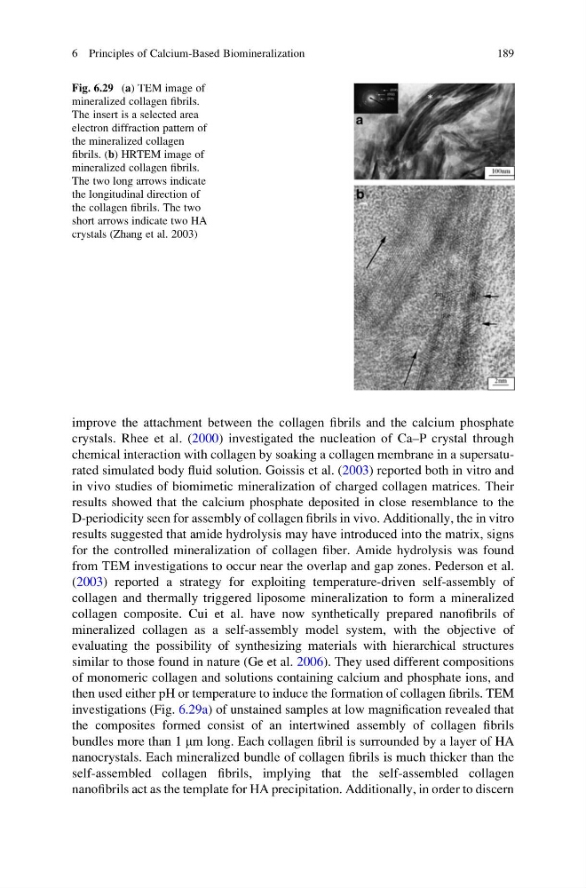
investigations (Fig. 6.29a) of unstained samples at low magnification revealed that
the composites formed consist of an intertwined assembly of collagen fibrils
bundles more than 1 mm long. Each collagen fibril is surrounded by a layer of HA
nanocrystals. Each mineralized bundle of collagen fibrils is much thicker than the
self-assembled collagen fibrils, implying that the self-assembled collagen
nanofibrils act as the template for HA precipitation. Additionally, in order to discern
Fig. 6.29 (a) TEM image of
mineralized collagen fibrils.
The insert is a selected area
electron diffraction pattern of
the mineralized collagen
fibrils. (b) HRTEM image of
mineralized collagen fibrils.
The two long arrows indicate
the longitudinal direction of
the collagen fibrils. The two
short arrows indicate two HA
crystals (Zhang et al. 2003)
6 Principles of Calcium-Based Biomineralization
189
crystals. Rhee et al. (2000) investigated the nucleation of Ca–P crystal through
chemical interaction with collagen by soaking a collagen membrane in a supersaturated simulated body fluid solution. Goissis et al. (2003) reported both in vitro and
in vivo studies of biomimetic mineralization of charged collagen matrices. Their
results showed that the calcium phosphate deposited in close resemblance to the
D-periodicity seen for assembly of collagen fibrils in vivo. Additionally, the in vitro
results suggested that amide hydrolysis may have introduced into the matrix, signs
for the controlled mineralization of collagen fiber. Amide hydrolysis was found
from TEM investigations to occur near the overlap and gap zones. Pederson et al.
(2003) reported a strategy for exploiting temperature-driven self-assembly of
collagen and thermally triggered liposome mineralization to form a mineralized
collagen composite. Cui et al. have now synthetically prepared nanofibrils of
mineralized collagen as a self-assembly model system, with the objective of
evaluating the possibility of synthesizing materials with hierarchical structures
similar to those found in nature (Ge et al. 2006). They used different compositions
of monomeric collagen and solutions containing calcium and phosphate ions, and
then used either pH or temperature to induce the formation of collagen fibrils. TEM
investigations (Fig. 6.29a) of unstained samples at low magnification revealed that
the composites formed consist of an intertwined assembly of collagen fibrils
bundles more than 1 mm long. Each collagen fibril is surrounded by a layer of HA
nanocrystals. Each mineralized bundle of collagen fibrils is much thicker than the
self-assembled collagen fibrils, implying that the self-assembled collagen
nanofibrils act as the template for HA precipitation. Additionally, in order to discern
Fig. 6.29 (a) TEM image of
mineralized collagen fibrils.
The insert is a selected area
electron diffraction pattern of
the mineralized collagen
fibrils. (b) HRTEM image of
mineralized collagen fibrils.
The two long arrows indicate
the longitudinal direction of
the collagen fibrils. The two
short arrows indicate two HA
crystals (Zhang et al. 2003)
6 Principles of Calcium-Based Biomineralization
189