

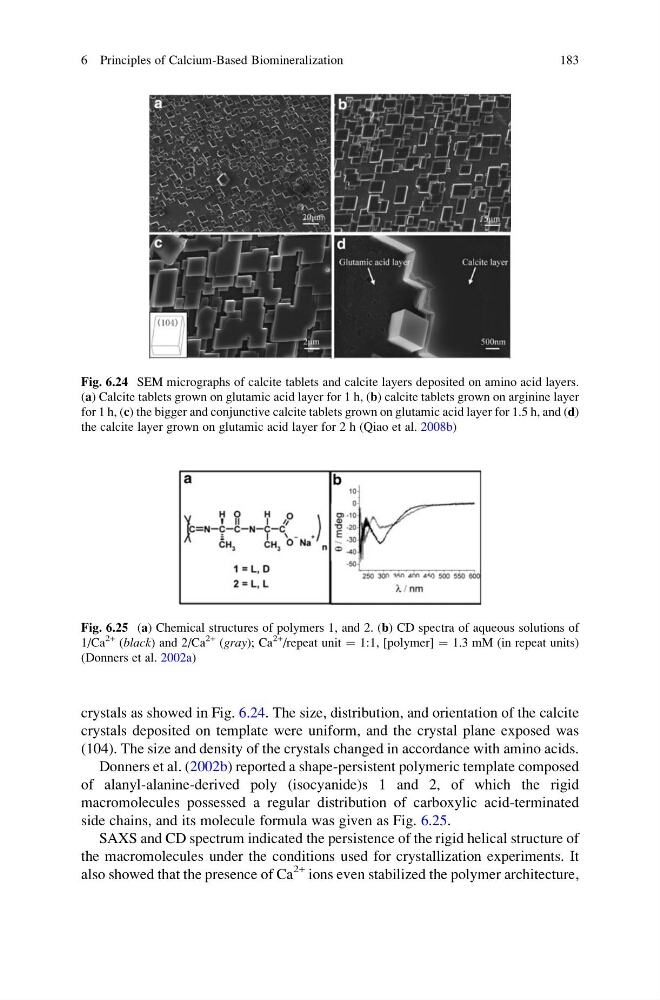
crystals as showed in Fig. 6.24. The size, distribution, and orientation of the calcite
crystals deposited on template were uniform, and the crystal plane exposed was
(104). The size and density of the crystals changed in accordance with amino acids.
Donners et al. (2002b) reported a shape-persistent polymeric template composed
of alanyl-alanine-derived poly (isocyanide)s 1 and 2, of which the rigid
macromolecules possessed a regular distribution of carboxylic acid-terminated
side chains, and its molecule formula was given as Fig. 6.25.
SAXS and CD spectrum indicated the persistence of the rigid helical structure of
the macromolecules under the conditions used for crystallization experiments. It
also showed that the presence of Ca
2+ ions even stabilized the polymer architecture,
Fig. 6.24 SEM micrographs of calcite tablets and calcite layers deposited on amino acid layers.
(a) Calcite tablets grown on glutamic acid layer for 1 h, (b) calcite tablets grown on arginine layer
for 1 h, (c) the bigger and conjunctive calcite tablets grown on glutamic acid layer for 1.5 h, and (d)
the calcite layer grown on glutamic acid layer for 2 h (Qiao et al. 2008b)
Fig. 6.25 (a) Chemical structures of polymers 1, and 2. (b) CD spectra of aqueous solutions of
1/Ca
2+ (black) and 2/Ca
2+ (gray); Ca
2+ /repeat unit ¼ 1:1, [polymer] ¼ 1.3 mM (in repeat units)
(Donners et al. 2002a)
6 Principles of Calcium-Based Biomineralization
183
crystals deposited on template were uniform, and the crystal plane exposed was
(104). The size and density of the crystals changed in accordance with amino acids.
Donners et al. (2002b) reported a shape-persistent polymeric template composed
of alanyl-alanine-derived poly (isocyanide)s 1 and 2, of which the rigid
macromolecules possessed a regular distribution of carboxylic acid-terminated
side chains, and its molecule formula was given as Fig. 6.25.
SAXS and CD spectrum indicated the persistence of the rigid helical structure of
the macromolecules under the conditions used for crystallization experiments. It
also showed that the presence of Ca
2+ ions even stabilized the polymer architecture,
Fig. 6.24 SEM micrographs of calcite tablets and calcite layers deposited on amino acid layers.
(a) Calcite tablets grown on glutamic acid layer for 1 h, (b) calcite tablets grown on arginine layer
for 1 h, (c) the bigger and conjunctive calcite tablets grown on glutamic acid layer for 1.5 h, and (d)
the calcite layer grown on glutamic acid layer for 2 h (Qiao et al. 2008b)
Fig. 6.25 (a) Chemical structures of polymers 1, and 2. (b) CD spectra of aqueous solutions of
1/Ca
2+ (black) and 2/Ca
2+ (gray); Ca
2+ /repeat unit ¼ 1:1, [polymer] ¼ 1.3 mM (in repeat units)
(Donners et al. 2002a)
6 Principles of Calcium-Based Biomineralization
183