

divisions inside the enamel crystals, determined by the two-stage mineralization
process. It can be presumed that the minerals in the fusion interfaces between the
nanofibrils or their small groups are vulnerable in acidic environments; thus, the
divisions of the crystals are eventually sculptured in the eroded surface. This is
because of the unique maturation stage, as well as the significant structural and
componential changes that enamel undergoes. An explanation of the formation of
the patterns observed in this study is complicated, and providing further direct
evidence is still a challenge for future studies.
One interesting characteristic of the hierarchical assembly to be highlighted is
that the scale distribution of the representative patterns at all levels appears regular,
indicating that the scale of each level is around ten times than that of the next
primary level. It is important to note that similar characteristics are found in the
structures of both human and fish bone (Wang et al. 2004). Taken together, these
results may imply an inherent regulation of mineralized hard connective tissues.
Moreover, such a regulation may exist not only in the natural biomaterials but also
in some self-assembly synthesized materials (Zhang et al. 2003a). Therefore, such
structural motifs presumably have the advantage of stabilizing systems that consist
of a large amount of small subunits. Although giving theoretical explanations for
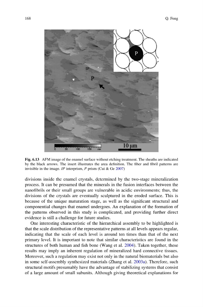
Fig. 6.13 AFM image of the enamel surface without etching treatment. The sheaths are indicated
by the black arrows. The insert illustrates the area definition. The fiber and fibril patterns are
invisible in the image. IP interprism, P prism (Cui & Ge 2007)
168
Q. Feng
process. It can be presumed that the minerals in the fusion interfaces between the
nanofibrils or their small groups are vulnerable in acidic environments; thus, the
divisions of the crystals are eventually sculptured in the eroded surface. This is
because of the unique maturation stage, as well as the significant structural and
componential changes that enamel undergoes. An explanation of the formation of
the patterns observed in this study is complicated, and providing further direct
evidence is still a challenge for future studies.
One interesting characteristic of the hierarchical assembly to be highlighted is
that the scale distribution of the representative patterns at all levels appears regular,
indicating that the scale of each level is around ten times than that of the next
primary level. It is important to note that similar characteristics are found in the
structures of both human and fish bone (Wang et al. 2004). Taken together, these
results may imply an inherent regulation of mineralized hard connective tissues.
Moreover, such a regulation may exist not only in the natural biomaterials but also
in some self-assembly synthesized materials (Zhang et al. 2003a). Therefore, such
structural motifs presumably have the advantage of stabilizing systems that consist
of a large amount of small subunits. Although giving theoretical explanations for
Fig. 6.13 AFM image of the enamel surface without etching treatment. The sheaths are indicated
by the black arrows. The insert illustrates the area definition. The fiber and fibril patterns are
invisible in the image. IP interprism, P prism (Cui & Ge 2007)
168
Q. Feng