

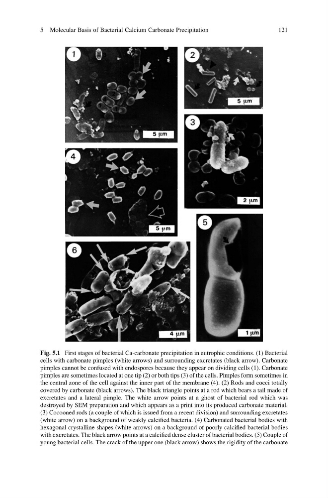
Fig. 5.1 First stages of bacterial Ca-carbonate precipitation in eutrophic conditions. (1) Bacterial
cells with carbonate pimples (white arrows) and surrounding excretates (black arrow). Carbonate
pimples cannot be confused with endospores because they appear on dividing cells (1). Carbonate
pimples are sometimes located at one tip (2) or both tips (3) of the cells. Pimples form sometimes in
the central zone of the cell against the inner part of the membrane (4). (2) Rods and cocci totally
covered by carbonate (black arrows). The black triangle points at a rod which bears a tail made of
excretates and a lateral pimple. The white arrow points at a ghost of bacterial rod which was
destroyed by SEM preparation and which appears as a print into its produced carbonate material.
(3) Cocooned rods (a couple of which is issued from a recent division) and surrounding excretates
(white arrow) on a background of weakly calcified bacteria. (4) Carbonated bacterial bodies with
hexagonal crystalline shapes (white arrows) on a background of poorly calcified bacterial bodies
with excretates. The black arrow points at a calcified dense cluster of bacterial bodies. (5) Couple of
young bacterial cells. The crack of the upper one (black arrow) shows the rigidity of the carbonate
5 Molecular Basis of Bacterial Calcium Carbonate Precipitation
121
cells with carbonate pimples (white arrows) and surrounding excretates (black arrow). Carbonate
pimples cannot be confused with endospores because they appear on dividing cells (1). Carbonate
pimples are sometimes located at one tip (2) or both tips (3) of the cells. Pimples form sometimes in
the central zone of the cell against the inner part of the membrane (4). (2) Rods and cocci totally
covered by carbonate (black arrows). The black triangle points at a rod which bears a tail made of
excretates and a lateral pimple. The white arrow points at a ghost of bacterial rod which was
destroyed by SEM preparation and which appears as a print into its produced carbonate material.
(3) Cocooned rods (a couple of which is issued from a recent division) and surrounding excretates
(white arrow) on a background of weakly calcified bacteria. (4) Carbonated bacterial bodies with
hexagonal crystalline shapes (white arrows) on a background of poorly calcified bacterial bodies
with excretates. The black arrow points at a calcified dense cluster of bacterial bodies. (5) Couple of
young bacterial cells. The crack of the upper one (black arrow) shows the rigidity of the carbonate
5 Molecular Basis of Bacterial Calcium Carbonate Precipitation
121