

be kept under controlled laboratory conditions for over 5 years (LePennec et al.
2003). Our data revealed that those Mn(II)-oxidizing bacteria are highly related to
the Mn(II)-oxidizing Bacillus strains isolated from Guaymas Basin, a deep-sea
hydrothermal vent environment in the Gulf of California (Dick et al. 2006).
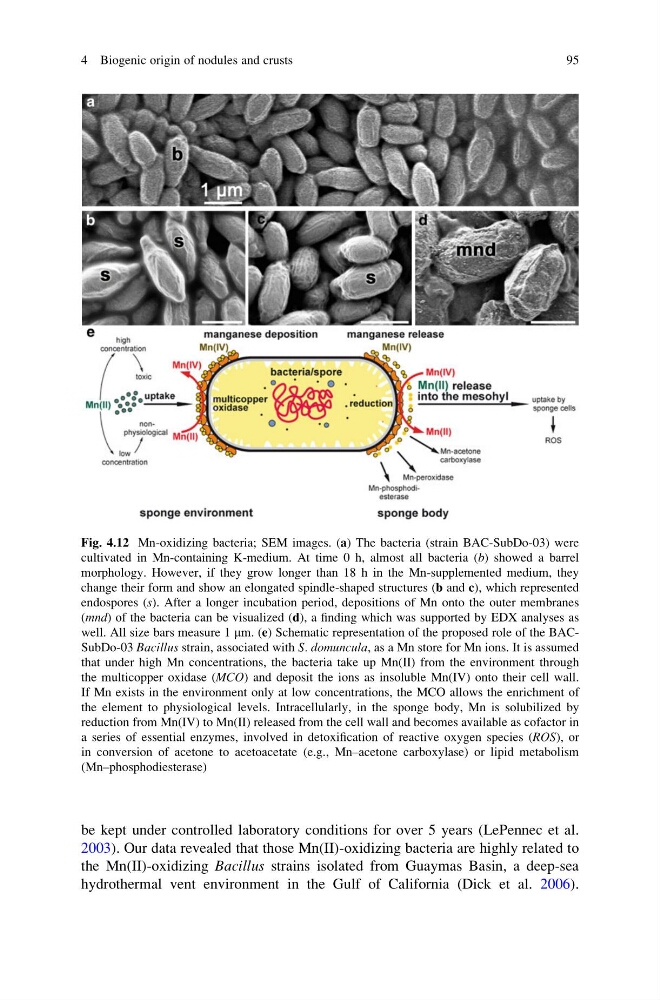
Fig. 4.12 Mn-oxidizing bacteria; SEM images. (a) The bacteria (strain BAC-SubDo-03) were
cultivated in Mn-containing K-medium. At time 0 h, almost all bacteria (b) showed a barrel
morphology. However, if they grow longer than 18 h in the Mn-supplemented medium, they
change their form and show an elongated spindle-shaped structures (b and c), which represented
endospores (s). After a longer incubation period, depositions of Mn onto the outer membranes
(mnd) of the bacteria can be visualized (d), a finding which was supported by EDX analyses as
well. All size bars measure 1 mm. (e) Schematic representation of the proposed role of the BACSubDo-03 Bacillus strain, associated with S. domuncula, as a Mn store for Mn ions. It is assumed
that under high Mn concentrations, the bacteria take up Mn(II) from the environment through
the multicopper oxidase (MCO) and deposit the ions as insoluble Mn(IV) onto their cell wall.
If Mn exists in the environment only at low concentrations, the MCO allows the enrichment of
the element to physiological levels. Intracellularly, in the sponge body, Mn is solubilized by
reduction from Mn(IV) to Mn(II) released from the cell wall and becomes available as cofactor in
a series of essential enzymes, involved in detoxification of reactive oxygen species (ROS), or
in conversion of acetone to acetoacetate (e.g., Mn–acetone carboxylase) or lipid metabolism
(Mn–phosphodiesterase)
4 Biogenic origin of nodules and crusts
95
2003). Our data revealed that those Mn(II)-oxidizing bacteria are highly related to
the Mn(II)-oxidizing Bacillus strains isolated from Guaymas Basin, a deep-sea
hydrothermal vent environment in the Gulf of California (Dick et al. 2006).
Fig. 4.12 Mn-oxidizing bacteria; SEM images. (a) The bacteria (strain BAC-SubDo-03) were
cultivated in Mn-containing K-medium. At time 0 h, almost all bacteria (b) showed a barrel
morphology. However, if they grow longer than 18 h in the Mn-supplemented medium, they
change their form and show an elongated spindle-shaped structures (b and c), which represented
endospores (s). After a longer incubation period, depositions of Mn onto the outer membranes
(mnd) of the bacteria can be visualized (d), a finding which was supported by EDX analyses as
well. All size bars measure 1 mm. (e) Schematic representation of the proposed role of the BACSubDo-03 Bacillus strain, associated with S. domuncula, as a Mn store for Mn ions. It is assumed
that under high Mn concentrations, the bacteria take up Mn(II) from the environment through
the multicopper oxidase (MCO) and deposit the ions as insoluble Mn(IV) onto their cell wall.
If Mn exists in the environment only at low concentrations, the MCO allows the enrichment of
the element to physiological levels. Intracellularly, in the sponge body, Mn is solubilized by
reduction from Mn(IV) to Mn(II) released from the cell wall and becomes available as cofactor in
a series of essential enzymes, involved in detoxification of reactive oxygen species (ROS), or
in conversion of acetone to acetoacetate (e.g., Mn–acetone carboxylase) or lipid metabolism
(Mn–phosphodiesterase)
4 Biogenic origin of nodules and crusts
95