

16S gene (Fig. 4.10) was found to share high sequence similarity (97%) to the
partial 16S ribosomal RNA gene from an uncultured bacterial clone JH-WH45
(AQbac_EF4928; accession number EF492894 [He et al. 2008]) with an E value
(maximal score) of 1,991. It is interesting to note that the bacteria that the authors
(He et al. 2008) described live in a Mn oxide-rich micro-biotope in soil environment. In the future, more sophisticated and more secure methods with respect to
contamination with environmental bacteria will have to be applied in order to draw
some conclusions on the possible etiology of modern bacteria with bacteria found
as fossils embedded in the minerals of the nodules.
4.4.9 Manganese Depositing Bacteria
As outlined above, the S-layer structures present on the surfaces of many bacteria
have been implicated in the nonenzymatic manganese deposition during nodule
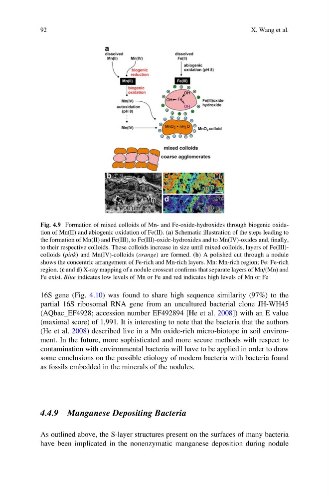
Fig. 4.9 Formation of mixed colloids of Mn- and Fe-oxide-hydroxides through biogenic oxidation of Mn(II) and abiogenic oxidation of Fe(II). (a) Schematic illustration of the steps leading to
the formation of Mn(II) and Fe(III), to Fe(III)-oxide-hydroxides and to Mn(IV)-oxides and, finally,
to their respective colloids. These colloids increase in size until mixed colloids, layers of Fe(III)colloids (pink) and Mn(IV)-colloids (orange) are formed. (b) A polished cut through a nodule
shows the concentric arrangement of Fe-rich and Mn-rich layers. Mn: Mn-rich region; Fe: Fe-rich
region. (c and d) X-ray mapping of a nodule crosscut confirms that separate layers of Mn/(Mn) and
Fe exist. Blue indicates low levels of Mn or Fe and red indicates high levels of Mn or Fe
92
X. Wang et al.
partial 16S ribosomal RNA gene from an uncultured bacterial clone JH-WH45
(AQbac_EF4928; accession number EF492894 [He et al. 2008]) with an E value
(maximal score) of 1,991. It is interesting to note that the bacteria that the authors
(He et al. 2008) described live in a Mn oxide-rich micro-biotope in soil environment. In the future, more sophisticated and more secure methods with respect to
contamination with environmental bacteria will have to be applied in order to draw
some conclusions on the possible etiology of modern bacteria with bacteria found
as fossils embedded in the minerals of the nodules.
4.4.9 Manganese Depositing Bacteria
As outlined above, the S-layer structures present on the surfaces of many bacteria
have been implicated in the nonenzymatic manganese deposition during nodule
Fig. 4.9 Formation of mixed colloids of Mn- and Fe-oxide-hydroxides through biogenic oxidation of Mn(II) and abiogenic oxidation of Fe(II). (a) Schematic illustration of the steps leading to
the formation of Mn(II) and Fe(III), to Fe(III)-oxide-hydroxides and to Mn(IV)-oxides and, finally,
to their respective colloids. These colloids increase in size until mixed colloids, layers of Fe(III)colloids (pink) and Mn(IV)-colloids (orange) are formed. (b) A polished cut through a nodule
shows the concentric arrangement of Fe-rich and Mn-rich layers. Mn: Mn-rich region; Fe: Fe-rich
region. (c and d) X-ray mapping of a nodule crosscut confirms that separate layers of Mn/(Mn) and
Fe exist. Blue indicates low levels of Mn or Fe and red indicates high levels of Mn or Fe
92
X. Wang et al.