

4.4.8 Approach to Determine Bacteria Species
in the Mineralic Material
It is very evident that isolation of DNA from micronodule material and the results
obtained after subsequent analyses of sequences from those samples must be taken
with greatest caution. For our first approach, pieces from a polymetallic nodule
were washed thoroughly with detergent and subsequently ground (Wang et al.
2009d). Then the material was leached with citric acid and the resulting material
subjected to DNA isolation followed by PCR analysis. By applying primers specific
for 16S ribosomal RNA gene, 28 clones were obtained after PCR reaction. Among
those, 19 sequences were identical; they comprised a 1,097-nucleotides-long 16S
ribosomal RNA gene sequence (it was termed: AQbac_NOD1_D). This partial
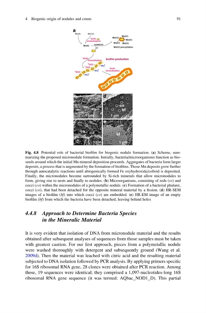
Fig. 4.8 Potential role of bacterial biofilm for biogenic nodule formation. (a) Scheme, summarizing the proposed micronodule formation. Initially, bacteria/microorganisms function as bioseeds around which the initial Mn mineral deposition proceeds. Aggregates of bacteria form larger
deposits, a process that is augmented by the formation of biofilms. Those Mn deposits grow further
through autocatalytic reactions until abiogenically formed Fe oxyhydroxide/colloid is deposited.
Finally, the micronodules become surrounded by Si-rich minerals that allow micronodules to
form, giving rise to nests and finally to nodules. (b) Microorganisms, consisting of rods (ro) and
cocci (co) within the micronodules of a polymetallic nodule. (c) Formation of a bacterial phalanx,
cocci (co), that had been detached for the opposite mineral material by a fission. (d) HR-SEM
images of a biofilm (bf) into which cocci (co) are embedded. (e) HR-EM image of an empty
biofilm (bf) from which the bacteria have been detached, leaving behind holes
4 Biogenic origin of nodules and crusts
91
in the Mineralic Material
It is very evident that isolation of DNA from micronodule material and the results
obtained after subsequent analyses of sequences from those samples must be taken
with greatest caution. For our first approach, pieces from a polymetallic nodule
were washed thoroughly with detergent and subsequently ground (Wang et al.
2009d). Then the material was leached with citric acid and the resulting material
subjected to DNA isolation followed by PCR analysis. By applying primers specific
for 16S ribosomal RNA gene, 28 clones were obtained after PCR reaction. Among
those, 19 sequences were identical; they comprised a 1,097-nucleotides-long 16S
ribosomal RNA gene sequence (it was termed: AQbac_NOD1_D). This partial
Fig. 4.8 Potential role of bacterial biofilm for biogenic nodule formation. (a) Scheme, summarizing the proposed micronodule formation. Initially, bacteria/microorganisms function as bioseeds around which the initial Mn mineral deposition proceeds. Aggregates of bacteria form larger
deposits, a process that is augmented by the formation of biofilms. Those Mn deposits grow further
through autocatalytic reactions until abiogenically formed Fe oxyhydroxide/colloid is deposited.
Finally, the micronodules become surrounded by Si-rich minerals that allow micronodules to
form, giving rise to nests and finally to nodules. (b) Microorganisms, consisting of rods (ro) and
cocci (co) within the micronodules of a polymetallic nodule. (c) Formation of a bacterial phalanx,
cocci (co), that had been detached for the opposite mineral material by a fission. (d) HR-SEM
images of a biofilm (bf) into which cocci (co) are embedded. (e) HR-EM image of an empty
biofilm (bf) from which the bacteria have been detached, leaving behind holes
4 Biogenic origin of nodules and crusts
91