

216
Ron H. Douglas
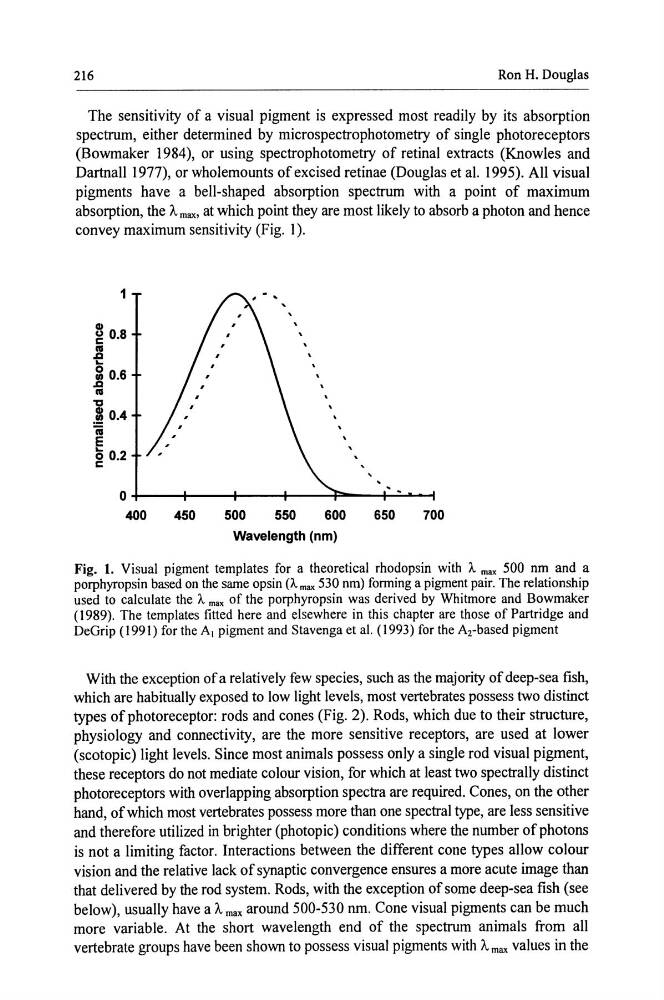
The sensitivity of a visual pigment is expressed most readily by its absorption
spectrum, either determined by microspectrophotometry of single photoreceptors
(Bowmaker 1984), or using spectrophotometry of retinal extracts (Knowles and
Dartnall 1977), or wholemounts of excised retinae (Douglas et al. 1995). All visual
pigments have a bell-shaped absorption spectrum with a point of maximum
absorption, the f.. max. at which point they are most likely to absorb a photon and hence
convey maximum sensitivity (Fig. 1 ).
1
CD
g 0.8
.e 0
1: 0.6
Ill
'0
10.4
ii
e g 0.2
0+----+----+----+----T----+~~
400
450
500
550
600
650
700
Wavelength (nm)
Fig. 1. Visual pigment templates for a theoretical rhodopsin with A. max 500 nm and a
porphyropsin based on the same opsin (A. max 530 nm) forming a pigment pair. The relationship
used to calculate the A. max of the porphyropsin was derived by Whitmore and Bowmaker
( 1989). The templates fitted here and elsewhere in this chapter are those of Partridge and
DeGrip ( 1991) for the A1 pigment and Stavenga et al. (1993) for the A2-based pigment
With the exception of a relatively few species, such as the majority of deep-sea fish,
which are habitually exposed to low light levels, most vertebrates possess two distinct
types of photoreceptor: rods and cones (Fig. 2). Rods, which due to their structure,
physiology and connectivity, are the more sensitive receptors, are used at lower
(scotopic) light levels. Since most animals possess only a single rod visual pigment,
these receptors do not mediate colour vision, for which at least two spectrally distinct
photoreceptors with overlapping absorption spectra are required. Cones, on the other
hand, of which most vertebrates possess more than one spectral type, are less sensitive
and therefore utilized in brighter (photopic) conditions where the number of photons
is not a limiting factor. Interactions between the different cone types allow colour
vision and the relative lack of synaptic convergence ensures a more acute image than
that delivered by the rod system. Rods, with the exception of some deep-sea fish (see
below), usually have a f.. max around 500-530 nm. Cone visual pigments can be much
more variable. At the short wavelength end of the spectrum animals from all
vertebrate groups have been shown to possess visual pigments with A max values in the
Ron H. Douglas
The sensitivity of a visual pigment is expressed most readily by its absorption
spectrum, either determined by microspectrophotometry of single photoreceptors
(Bowmaker 1984), or using spectrophotometry of retinal extracts (Knowles and
Dartnall 1977), or wholemounts of excised retinae (Douglas et al. 1995). All visual
pigments have a bell-shaped absorption spectrum with a point of maximum
absorption, the f.. max. at which point they are most likely to absorb a photon and hence
convey maximum sensitivity (Fig. 1 ).
1
CD
g 0.8
.e 0
1: 0.6
Ill
'0
10.4
ii
e g 0.2
0+----+----+----+----T----+~~
400
450
500
550
600
650
700
Wavelength (nm)
Fig. 1. Visual pigment templates for a theoretical rhodopsin with A. max 500 nm and a
porphyropsin based on the same opsin (A. max 530 nm) forming a pigment pair. The relationship
used to calculate the A. max of the porphyropsin was derived by Whitmore and Bowmaker
( 1989). The templates fitted here and elsewhere in this chapter are those of Partridge and
DeGrip ( 1991) for the A1 pigment and Stavenga et al. (1993) for the A2-based pigment
With the exception of a relatively few species, such as the majority of deep-sea fish,
which are habitually exposed to low light levels, most vertebrates possess two distinct
types of photoreceptor: rods and cones (Fig. 2). Rods, which due to their structure,
physiology and connectivity, are the more sensitive receptors, are used at lower
(scotopic) light levels. Since most animals possess only a single rod visual pigment,
these receptors do not mediate colour vision, for which at least two spectrally distinct
photoreceptors with overlapping absorption spectra are required. Cones, on the other
hand, of which most vertebrates possess more than one spectral type, are less sensitive
and therefore utilized in brighter (photopic) conditions where the number of photons
is not a limiting factor. Interactions between the different cone types allow colour
vision and the relative lack of synaptic convergence ensures a more acute image than
that delivered by the rod system. Rods, with the exception of some deep-sea fish (see
below), usually have a f.. max around 500-530 nm. Cone visual pigments can be much
more variable. At the short wavelength end of the spectrum animals from all
vertebrate groups have been shown to possess visual pigments with A max values in the