

The Design of Compound Eyes and the Illumination of Natural Habitats
189
All compound eyes are composed of tube-like optical units called ommatidia.
Each ommatidium consists of a lens element (the cornea and crystalline cone) that
focuses light incident from a narrow region of space onto an underlying bundle of
photoreceptors (the rhabdom). A compound eye may contain as few as 6 ommatidia,
as in some ants, or as many as 30 000 as in large dragonflies. The number of ommatidia
is usually a good indicator of the spatial resolution of a particular eye. The greater
the number of ommatidia, and the denser their packing, the finer the resolution of
the eye. In fact, the angle between neighbouring ommatidia - the interommatidial
angle, .1 eye with an interommatidial angle of0.24° (as in the dorsal eyes of some dragonflies:
Sherk 1978) has greater anatomical resolution than an eye with an interommatidial
angle of 4° (as might be found in a ground beetle). To put this into perspective, the
angle between the cones of our fovea is around 0.0 l o, so our anatomical resolution is
significantly greater! The interommatidial angles of various compound eyes are given
in Table 1.
Apposition Compound Eye
Superposition Compound Eye
p
d
A
B
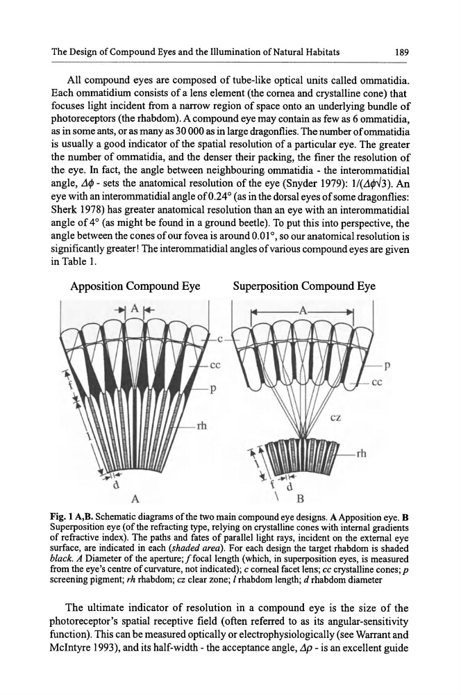
Fig. 1 A,B. Schematic diagrams of the two main compound eye designs. A Apposition eye. B
Superposition eye (of the refracting type, relying on crystalline cones with internal gradients
of refractive index). The paths and fates of parallel light rays, incident on the external eye
surface, are indicated in each (shaded area). For each design the target rhabdom is shaded
black. A Diameter of the aperture;! focal length (which, in superposition eyes, is measured
from the eye's centre of curvature, not indicated); c corneal facet lens; cc crystalline cones; p
screening pigment; rh rhabdom; cz clear zone; l rhabdom length; d rhabdom diameter
The ultimate indicator of resolution in a compound eye is the size of the
photoreceptor's spatial receptive field (often referred to as its angular-sensitivity
function). This can be measured optically or electrophysiologically (see Warrant and
Mcintyre 1993), and its half-width- the acceptance angle, L1p- is an excellent guide
189
All compound eyes are composed of tube-like optical units called ommatidia.
Each ommatidium consists of a lens element (the cornea and crystalline cone) that
focuses light incident from a narrow region of space onto an underlying bundle of
photoreceptors (the rhabdom). A compound eye may contain as few as 6 ommatidia,
as in some ants, or as many as 30 000 as in large dragonflies. The number of ommatidia
is usually a good indicator of the spatial resolution of a particular eye. The greater
the number of ommatidia, and the denser their packing, the finer the resolution of
the eye. In fact, the angle between neighbouring ommatidia - the interommatidial
angle, .1 eye with an interommatidial angle of0.24° (as in the dorsal eyes of some dragonflies:
Sherk 1978) has greater anatomical resolution than an eye with an interommatidial
angle of 4° (as might be found in a ground beetle). To put this into perspective, the
angle between the cones of our fovea is around 0.0 l o, so our anatomical resolution is
significantly greater! The interommatidial angles of various compound eyes are given
in Table 1.
Apposition Compound Eye
Superposition Compound Eye
p
d
A
B
Fig. 1 A,B. Schematic diagrams of the two main compound eye designs. A Apposition eye. B
Superposition eye (of the refracting type, relying on crystalline cones with internal gradients
of refractive index). The paths and fates of parallel light rays, incident on the external eye
surface, are indicated in each (shaded area). For each design the target rhabdom is shaded
black. A Diameter of the aperture;! focal length (which, in superposition eyes, is measured
from the eye's centre of curvature, not indicated); c corneal facet lens; cc crystalline cones; p
screening pigment; rh rhabdom; cz clear zone; l rhabdom length; d rhabdom diameter
The ultimate indicator of resolution in a compound eye is the size of the
photoreceptor's spatial receptive field (often referred to as its angular-sensitivity
function). This can be measured optically or electrophysiologically (see Warrant and
Mcintyre 1993), and its half-width- the acceptance angle, L1p- is an excellent guide