

151
The important subsequent step is defining our axes.
Here we
desire maximum separation of the various chromatic groups of cells.
After considerable experimentation we have settled on the use of DUN
(Dunatietta tertioteata, a chlorophyte) and DC-2 (Syneahoaoaaus sp.,
a phycoerythrin-containing cyanobacteria) grown under standard light,
temperature and transfer conditions,as our biological standards/
controls.
This permits us to construct a defined map.
Our map
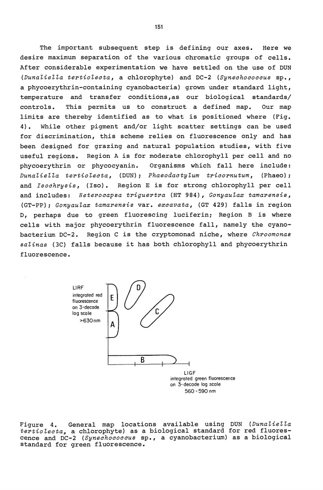
limits are thereby identified as to what is positioned where (Fig.
4) • While other pigment and/or light scatter settings can be used
for discrimination, this scheme relies on fluorescence only and has
been designed for grazing and natural population studies, with five
useful regions. Region A is for moderate chlorophyll per cell and no
phycoerythrin or phycocyanin.
DunaHeHa tel'tioteata, (DUN);
Organisms which fall here include:
Phaeodaatytum tl'iaol'nutum, (Phaeo);
and Isoahrysis, (Iso). Region E is for strong chlorophyll per cell
and includes: Hetel'oaapsa tl'iguel'tl'a (HT 984), Gonyautax tamal'ensis,
(GT-PP); Gonyautax tamarensis var. exaavata, (GT 429) falls in region
D, perhaps due to green fluorescing luciferin; Region B is where
cells with major phycoerythrin fluorescence fall, namely the cyanobacterium DC-2. Region C is the cryptomonad niche, where Chroomonas
satinas (3C) falls because it has both chlorophyll and phycoerythrin
fluorescence.
LlRF
integrated red
fl uarescence
an 3-decade
log scale
>630nm
B
LlGF
integrated green fluorescence
on 3- decade lag scale
560-590 nm
Figure 4.
General map locations available using DUN (DunaHeHa
tertioteata. a chlorophyte) as a biological standard for red fluorescence and DC-2 (Syneahoaoaaus sp., a cyanobacterium) as a biological
standard for green fluorescence.
The important subsequent step is defining our axes.
Here we
desire maximum separation of the various chromatic groups of cells.
After considerable experimentation we have settled on the use of DUN
(Dunatietta tertioteata, a chlorophyte) and DC-2 (Syneahoaoaaus sp.,
a phycoerythrin-containing cyanobacteria) grown under standard light,
temperature and transfer conditions,as our biological standards/
controls.
This permits us to construct a defined map.
Our map
limits are thereby identified as to what is positioned where (Fig.
4) • While other pigment and/or light scatter settings can be used
for discrimination, this scheme relies on fluorescence only and has
been designed for grazing and natural population studies, with five
useful regions. Region A is for moderate chlorophyll per cell and no
phycoerythrin or phycocyanin.
DunaHeHa tel'tioteata, (DUN);
Organisms which fall here include:
Phaeodaatytum tl'iaol'nutum, (Phaeo);
and Isoahrysis, (Iso). Region E is for strong chlorophyll per cell
and includes: Hetel'oaapsa tl'iguel'tl'a (HT 984), Gonyautax tamal'ensis,
(GT-PP); Gonyautax tamarensis var. exaavata, (GT 429) falls in region
D, perhaps due to green fluorescing luciferin; Region B is where
cells with major phycoerythrin fluorescence fall, namely the cyanobacterium DC-2. Region C is the cryptomonad niche, where Chroomonas
satinas (3C) falls because it has both chlorophyll and phycoerythrin
fluorescence.
LlRF
integrated red
fl uarescence
an 3-decade
log scale
>630nm
B
LlGF
integrated green fluorescence
on 3- decade lag scale
560-590 nm
Figure 4.
General map locations available using DUN (DunaHeHa
tertioteata. a chlorophyte) as a biological standard for red fluorescence and DC-2 (Syneahoaoaaus sp., a cyanobacterium) as a biological
standard for green fluorescence.