

Remote Sensing of Terrestrial Photosynthesis
521
24.4.2 Xanthophyll Pigments
The carotenoid pigments that comprise the xanthophyll cycle are clearly
implicated in the dissipation of absorbed PAR not used in photosynthesis
(Bjorkman and Demmig-Adams, Chap. 2, this Vol.). The total pool of
xanthophyll pigments increases with growth PAR. The quantity of pigment
in the de-epoxidized (energy-dissipating) form increases strongly as nonphotochemical quenching increases (Bjorkman and Demmig-Adams, this
Vol.) and as the realized photon efficiency (02 evolution or CO2 uptake
per unit of absorbed PAR) decreases (Demmig-Adams and Adams 1992;
Thayer and Bjorkman 1990). Remote sensing of the status of the xanthophyll pigments could lead to remote estimates of in situ photosynthetic
efficiency, which, in combination with estimates of downwelling PAR, could
lead to a new approach for estimating photosynthetic CO2 uptake.
Bilger et al. (1989) first reported that the status of the xanthophyll cycle
pigments influences leaf spectral absorptance. Following sudden illumination,
changes in leaf spectral absorptance in the green region represent the combined effect of redistribution of the xanthophyll pigments, plus changes
in light scattering resulting from chloroplast movements and changes in
thylakoid conformation (Brugnoli and Bjorkman 1992).
Using sunflower canopies, Gamon et al. (1990) observed two regions
of unstable spectral reflectance following sudden illumination. One region
(from about 670 to 800 nm) had the spectral characteristics of chlorophyll
fluorescence, while the other (from about 500 to 560 nm) had a wavelength
range expected for changes due to the interconversion of the xanthophyll
pigments and chloroplast movements. A strong correlation between the
reflectance at 531 nm and the epoxidation state of the xanthophyll pigments
in upper canopy leaves, as well as the elimination of the spectral instability
in the green in leaves fed dithiothreitol (an inhibitor of the deepoxidation
reaction responsible for the production of the energy dissipating xanthophyll
pigment), confirmed the involvement of the xanthophyll cycle pigments in
the spectral instability.
Gamon et al. (1992) considered approaches for quantifying the status
of the xanthophyll pigments without canopy-scale PAR manipulations. A
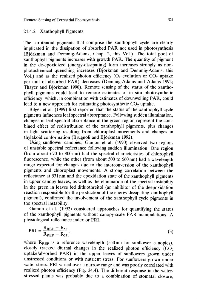
physiological reflectance index or PRI,
PRI = RREF - R531
RREF + RS31 '
(3)
where RREF is a reference wavelength (550nm for sunflower canopies),
closely tracked diurnal changes in the realized photon efficiency (C0 2
uptake/absorbed PAR) in the upper leaves of sunflowers grown under
unstressed conditions or with nutrient stress. For sunflowers grown under
water stress, PRI varied over a narrow range and was poorly correlated with
realized photon efficiency (Fig. 24.4). The different response in the waterstressed plants was probably due to a combination of stomatal closure,
521
24.4.2 Xanthophyll Pigments
The carotenoid pigments that comprise the xanthophyll cycle are clearly
implicated in the dissipation of absorbed PAR not used in photosynthesis
(Bjorkman and Demmig-Adams, Chap. 2, this Vol.). The total pool of
xanthophyll pigments increases with growth PAR. The quantity of pigment
in the de-epoxidized (energy-dissipating) form increases strongly as nonphotochemical quenching increases (Bjorkman and Demmig-Adams, this
Vol.) and as the realized photon efficiency (02 evolution or CO2 uptake
per unit of absorbed PAR) decreases (Demmig-Adams and Adams 1992;
Thayer and Bjorkman 1990). Remote sensing of the status of the xanthophyll pigments could lead to remote estimates of in situ photosynthetic
efficiency, which, in combination with estimates of downwelling PAR, could
lead to a new approach for estimating photosynthetic CO2 uptake.
Bilger et al. (1989) first reported that the status of the xanthophyll cycle
pigments influences leaf spectral absorptance. Following sudden illumination,
changes in leaf spectral absorptance in the green region represent the combined effect of redistribution of the xanthophyll pigments, plus changes
in light scattering resulting from chloroplast movements and changes in
thylakoid conformation (Brugnoli and Bjorkman 1992).
Using sunflower canopies, Gamon et al. (1990) observed two regions
of unstable spectral reflectance following sudden illumination. One region
(from about 670 to 800 nm) had the spectral characteristics of chlorophyll
fluorescence, while the other (from about 500 to 560 nm) had a wavelength
range expected for changes due to the interconversion of the xanthophyll
pigments and chloroplast movements. A strong correlation between the
reflectance at 531 nm and the epoxidation state of the xanthophyll pigments
in upper canopy leaves, as well as the elimination of the spectral instability
in the green in leaves fed dithiothreitol (an inhibitor of the deepoxidation
reaction responsible for the production of the energy dissipating xanthophyll
pigment), confirmed the involvement of the xanthophyll cycle pigments in
the spectral instability.
Gamon et al. (1992) considered approaches for quantifying the status
of the xanthophyll pigments without canopy-scale PAR manipulations. A
physiological reflectance index or PRI,
PRI = RREF - R531
RREF + RS31 '
(3)
where RREF is a reference wavelength (550nm for sunflower canopies),
closely tracked diurnal changes in the realized photon efficiency (C0 2
uptake/absorbed PAR) in the upper leaves of sunflowers grown under
unstressed conditions or with nutrient stress. For sunflowers grown under
water stress, PRI varied over a narrow range and was poorly correlated with
realized photon efficiency (Fig. 24.4). The different response in the waterstressed plants was probably due to a combination of stomatal closure,