

B
Pathological Effects
A
47
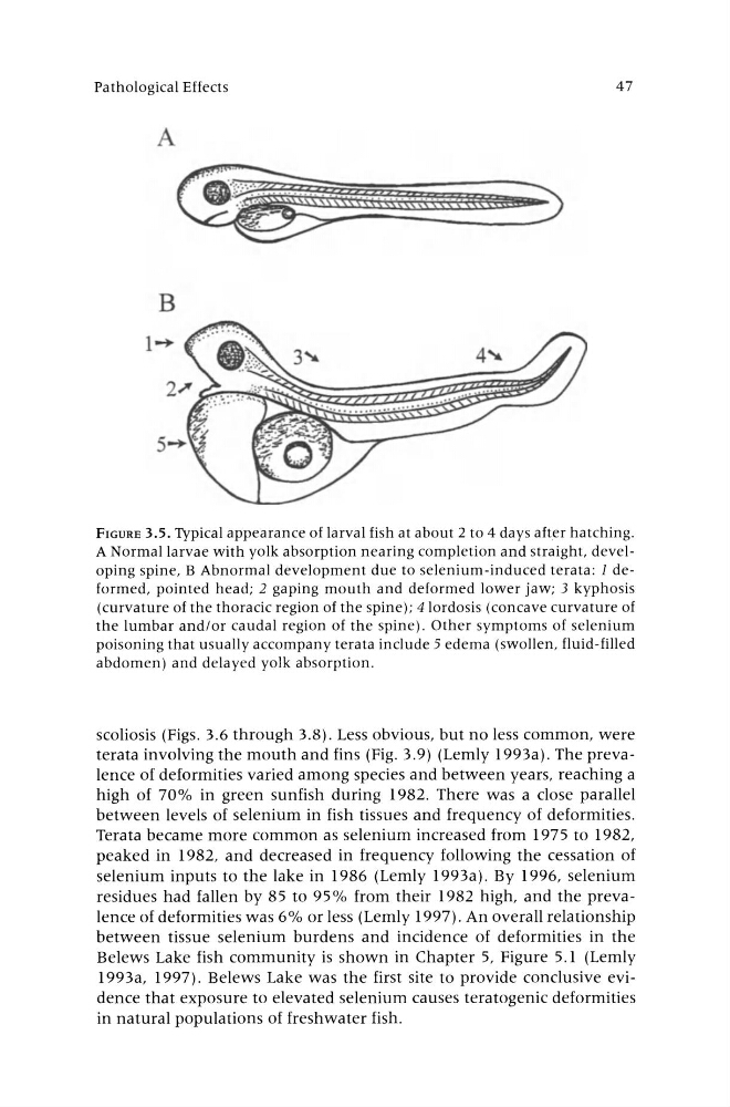
FIGURE 3.5. Typical appearance of larval fish at about 2 to 4 days after hatching.
A Normal larvae with yolk absorption nearing completion and straight. developing spine, B Abnormal development due to selenium-induced terata: 1 deformed, pointed head; 2 gaping mouth and deformed lower jaw; 3 kyphosis
(curvature of the thoracic region of the spine); 4 lordosis (concave curvature of
the lumbar and/or caudal region of the spine). Other symptoms of selenium
poisoning that usually accompany terata include 5 edema (swollen, fluid -filled
abdomen) and delayed yolk absorption.
scoliosis (Figs. 3.6 through 3.8). Less obvious, but no less common, were
terata involving the mouth and fins (Fig. 3.9) (Lemly 1993a). The prevalence of deformities varied among species and between years, reaching a
high of 70% in green sunfish during 1982. There was a close parallel
between levels of selenium in fish tissues and frequency of deformities.
Terata became more common as selenium increased from 1975 to 1982,
peaked in 1982, and decreased in frequency following the cessation of
selenium inputs to the lake in 1986 (Lemly 1993a). By 1996, selenium
residues had fallen by 85 to 95% from their 1982 high, and the prevalence of deformities was 6% or less (Lemly 1997). An overall relationship
between tissue selenium burdens and incidence of deformities in the
Belews Lake fish community is shown in Chapter 5, Figure 5.1 (Lemly
1993a, 1997). Belews Lake was the first site to provide conclusive evidence that exposure to elevated selenium causes teratogenic deformities
in natural populations of freshwater fish.
Pathological Effects
A
47
FIGURE 3.5. Typical appearance of larval fish at about 2 to 4 days after hatching.
A Normal larvae with yolk absorption nearing completion and straight. developing spine, B Abnormal development due to selenium-induced terata: 1 deformed, pointed head; 2 gaping mouth and deformed lower jaw; 3 kyphosis
(curvature of the thoracic region of the spine); 4 lordosis (concave curvature of
the lumbar and/or caudal region of the spine). Other symptoms of selenium
poisoning that usually accompany terata include 5 edema (swollen, fluid -filled
abdomen) and delayed yolk absorption.
scoliosis (Figs. 3.6 through 3.8). Less obvious, but no less common, were
terata involving the mouth and fins (Fig. 3.9) (Lemly 1993a). The prevalence of deformities varied among species and between years, reaching a
high of 70% in green sunfish during 1982. There was a close parallel
between levels of selenium in fish tissues and frequency of deformities.
Terata became more common as selenium increased from 1975 to 1982,
peaked in 1982, and decreased in frequency following the cessation of
selenium inputs to the lake in 1986 (Lemly 1993a). By 1996, selenium
residues had fallen by 85 to 95% from their 1982 high, and the prevalence of deformities was 6% or less (Lemly 1997). An overall relationship
between tissue selenium burdens and incidence of deformities in the
Belews Lake fish community is shown in Chapter 5, Figure 5.1 (Lemly
1993a, 1997). Belews Lake was the first site to provide conclusive evidence that exposure to elevated selenium causes teratogenic deformities
in natural populations of freshwater fish.